


")
")
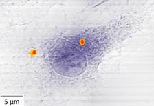
Two agglomerates of antibiotic-loaded iron nanocontainers (red) in a macrophage. Credit: Stachnik et al., „Scientific Reports“, CC BY 4.0 [Source]
")
")
")
")
With an advanced X-ray combination technique at PETRA III, scientists have traced nanocarriers for tuberculosis drugs within cells with very high precision. The method combines two sophisticated scanning X-ray measurements and can locate minute amounts of various metals in biological samples at very high resolution, as a team around DESY scientist Karolina Stachnik reports in the journal Scientific Reports. To illustrate its versatility, the researchers have also used the combination method to map the calcium content in human bone, an analysis that can benefit osteoporosis research.
“Metals play key roles in numerous biological processes, from the oxygen transport in our red blood cells and the mineralisation of bones to the detrimental accumulation of metals in nerve cells as seen in diseases like Alzheimer's,” explains Stachnik who works in the Center for Free-Electron Laser Science CFEL at DESY. High-energy X-rays make metals light up in fluorescence, a method that is very sensitive even to tiny amounts. “However, the X-ray fluorescence measurements usually do not show the ultrastructure of a cell, for example,” says DESY scientist Alke Meents who led the research. “If you want to exactly locate the metals within your sample, you have to combine the measurements with an imaging technique.” The ultrastructure comprises the details of the cell morphology that are not visible under an optical microscope.
As biological samples, such as cells, are very sensitive to X-ray radiation, it is highly beneficial to image their structure simultaneously to the fluorescence analysis. For this reason, the team combined the fluorescence measurements with an imaging method known as ptychography. “A ptychographic microscope is fairly similar to taking a panorama image,” explains Stachnik. “An extended specimen like a biological cell is raster scanned with a small coherent X-ray beam that produces many overlapping images of parts of the sample. These overlapping images are then stitched together afterwards.”
The applied method works without any lenses between the sample and the detector, and as a consequence so-called X-ray diffraction patterns are recorded on the detector. Each of these patterns contains information on the spatial structure of the respective part of the sample, which can be calculated from the pattern. “This finally results in a fully quantitative optical density map of the specimen”, explains Stachnik. “Via this complex process, ptychography provides spatial resolutions beyond the usual limits of X-ray optics.”
Thanks to its scanning nature, ptychography can be combined with simultaneous acquisition of X-ray fluorescence measurements that provide a unique fingerprint of the sample-constituting elements. In this way, a photograph of the sample's morphology obtained by ptychography can be overlaid with an element map. “The concurrent combination of these two complementary imaging methods enables therefore artefact-free correlations of trace elements with the highly resolved specimen’s structure,” summarises Meents.
A fundamental prerequisite is that the X-rays are of a single colour only (monochromatic, all having the same wavelength) and that they oscillate in step (coherent) like in a laser. “Sufficiently bright coherent monochromatic X-rays with energies high enough to let metals like iron fluoresce have only become available at modern synchrotron light sources like DESY's PETRA III”, says Meents.
To test the method, the DESY researchers teamed up with the group of Ulrich Schaible from the Research Center Borstel to investigate localisation and concentration of nanocarriers for tuberculosis drugs within macrophages, the scavenger cells of the immune system. “Usually, macrophages destroy pathogens like viruses and bacteria. Unfortunately, tuberculosis bacteria have managed to evade destruction and hide inside the macrophages instead, even using them to grow”, says Schaible. “As a barrier for effective treatment, the bacteria’s niches within macrophages need to be reached by antibiotics to be efficient.”
A new “Trojan Horse” strategy uses nanometre-sized iron containers to deliver antibiotics directly into the cells. These containers are hollow, filled with antibiotics and measure less than 20 nanometres in diameter (a nanometre is a millionth of a millimetre). “Macrophages swallow the containers, and once they are inside the cell, the iron walls of the cages slowly dissolve due to the need of the bacteria for iron. Eventually, the antibiotics are released and kill the bacteria”, explains Schaible.
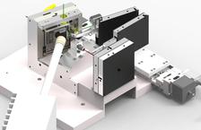
To evaluate the efficacy of this strategy, the team investigated macrophages that had been fed iron containers. Using a specially developed scanning stage at the bio-imaging and diffraction beamline P11 of DESY's X-ray source PETRA III, the researchers could capture ptychographic and fluorescence images of 14 cells with subcellular resolution and identified a total of 22 agglomerates of nanocontainers within them.
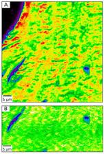
In a second application the researchers teamed up with the group of Björn Busse from the University Medical Center Hamburg-Eppendorf (UKE) and analysed the calcium content in a sample of human bone. “Calcium is a key element that makes our bones strong”, explains co-author Katharina Jähn from Busse's group. “However, in times of high calcium requirement, the body dissolves it from the bones to be used elsewhere. These and other age-related processes can lead to osteoporosis, affecting nearly a quarter of all women at ages over 50 years in Germany.”
Experimental research on bone mineralisation is usually performed on small slices of bone. “However, only the total content of calcium is usually mapped this way,” says Stachnik. “To get a true measure of the calcium concentration, one has to correct for the often varying thickness of the sample.” The team used a simultaneously obtained ptychographic image to remove the mass-thickness distortion from the calcium distribution map. “With this approach we were able to observe a locally lower calcium content at certain points in the bone, which helps to better understand the process of bone disorders and to quantify the effect of bone mineralisation changes in patients”, emphasises Stachnik.
To improve the method even further, the researchers have started to extend the analysis to three-dimensional measurements. “The experimental setup is currently being extended to allow acquisition of 3D-tomographic datasets at beamline P11,” says Meents. “With many synchrotrons being upgraded to produce even brighter X-rays, we expect the method to increase throughput and to become a routine application at these facilities.”
The Research Center Borstel, the Paul Scherrer Institute in Switzerland, the Karlsruhe Institute of Technology, the University Medical Center Hamburg-Eppendorf and DESY were involved in this research.
(from DESY News)
Reference:
Multimodal X-ray imaging of nanocontainer-treated macrophages and calcium distribution in the perilacunar bone matrix; Karolina Stachnik, Martin Warmer, Istvan Mohacsi, Vincent Hennicke, Pontus Fischer, Jan Meyer, Tobias Spitzbart, Miriam Barthelmess, Jacqueline Eich, Christian David, Claus Feldmann, Björn Busse, Katharina Jähn, Ulrich E. Schaible, and Alke Meents; Scientific Reports, 2020; DOI: 10.1038/s41598-020-58318-7