


")
")
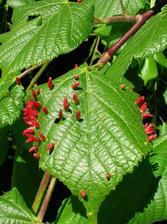
Nail galls on a leaf (Credit: Wikimedia Comms).
")
")
They can be a nightmare for farmers and gardeners: galls, abnormal plant growths caused by various organisms, can damage crops and other plants. Researchers have used PETRA III to analyse galls caused by mites that infest the leaves of various trees and bushes. What they found is incredible: The mites alter the plants’ metabolism of trace metals in the newly formed galls to better suit their needs, aggregating metals needed for their nutrition and protection while sequestering the excessive ones. The results, published in the journal New Phytologist, could help scientists to better understand these evolutionarily fascinating interactions between plants and specialised herbivores and perhaps eventually help avoid damage to economically important plants.
A gall can be formed on every part of a plant: leaf, root, trunk, flower. Insects and mites can induce complex galls by hijacking the plant’s cellular functions and extensively reprogramming expression of its genes. The new tissue serves to feed and protect the gall-inducing insect or mite. The mites from the family Eriophyidae make brightly coloured, spike-shaped “nail” galls, within which they spend multiple generations from spring to autumn each year. A female mite infests the emerging leaves in springtime, and the leaf grows with galls filled with numerous male and female mites. During winter, the female mites emerging from the galls hide in winter buds to initiate another life cycle in the spring.
“The gall is a sort of ordered tumour,” says Filis Morina, a plant scientist based at the Biology Centre of the Czech Academy of Sciences in České Budĕjovice who is first author of the study. “This is like a hijacking of the plant’s cellular functions because the mite completely manipulates what happens to the leaf. The leaf otherwise would never make such a shape as a nail gall.”
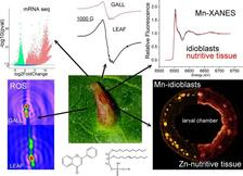
Although they are essential for both the plant and the mites, the role of the metals in the interaction between the two had remained elusive. To reveal this, the authors used a technique called tomography – more commonly used in medical imaging – and the X-rays of the DESY X-ray light source PETRA III to peer into the gall.
“In scanning X-ray fluorescence tomography, you scan the sample line by line, and you rotate the sample between the lines,” says DESY scientist Gerald Falkenberg, who leads the PETRA III P06 beamline and performed the research with the team. The result is a three-dimensional see-through map of the structures and contents of the galls.
“The P06 beamline at PETRA III allowed us to look down with subcellular detail as to where elements that are decisive for the development of the mite and the gall are distributed,” says Hendrik Küpper, who led the research with Morina at the Biology Centre CAS. The team found that mites induce accumulation of zinc, iron and copper in the nutritive tissue, while manganese and calcium accumulate in large secretory cells. The mites had changed the distribution of metals as well as their function. Using complementary spectroscopic, biochemical and genetic techniques, the team could finally map different metals with gene expression, metabolic profile and oxidation–reduction reactions regulated by the mite in the gall.
To further understand the changes in manganese metabolism, the team used a technique called XANES (X-ray absorption near edge spectroscopy) tomography to get a map of manganese absorption spectra in different gall compartments. This allowed for identification and mapping of the classes of manganese-binding compounds, linking distribution of the metal and its function in individual cells in the galls.
“Galls affect a great number of species, so you have crops, ornamental plants and economically important species affected,” says Morina. “Understanding how micronutrients change in these types of interactions, can be used in agriculture or forestry and even in urban ecosystems to protect plants from this kind of parasitism. It’s only the beginning of this type of research.”
PETRA IV, the planned 4-D X-ray microscope at DESY, will also make it possible to gain even more detailed insights into issues relating to leaf galls thanks to its high brilliance. Gerald Falkenberg explains: "The PETRA IV infrastructure can greatly contribute to the mechanistic understanding of the interactions between plants and gall-inducing organisms. With increased resolution at the CryoBioNanoprobe beamline at PETRA IV, subcellular distribution and speciation of metals in the cells with different functions in the plant galls (nutrition vs protection) and the galler itself (feeding apparatus, gut, ovipositors) will be revealed."
(from DESY News)
Reference:
F Morina et al, How eriophyd mites shape metal metabolism in leaf galls on Tilia cordata, New Phytologist (2025), DOI: 10.1111/nph.70103