


")
")
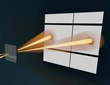
The sample (left) is scanned by two deflected X-ray beams (coming from the left) and both diffraction patterns are captured by the detector on the right. (Credit: Figure adopted from original publication)
")
")
Imagine being able to see the fine, three-dimensional (3D) structure of objects a hundred times thinner than a human hair, not just as flat images but in perspective—with depth and detail. In the joint project 'XStereoVision' a team of researchers from DESY, University of Hamburg and KTH Royal Institute of Technology (Sweden) have now made this possible with the newly developed stereo hard X-ray ptychography technique.
Understanding the internal structure of materials at the nanoscale is crucial for science and in particular for technological developments. Hard X-rays can penetrate deeply into materials and reveal fine details, but most traditional methods provide only flat, two-dimensional images. To see structures in 3D, typically tomography is used—like a medical computed tomography (CT) scan—which requires several measurements and is often time consuming. Ptychography offers high-resolution imaging by scanning a sample with a spatially confined and coherent X-ray beam. However, to obtain a 3D impression with this powerful technique, the sample had to be rotated until now.
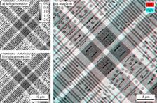
In their new approach, the researchers took inspiration from how our two eyes enable depth perception. They collected two ptychographic images of the same sample from slightly different angles. By combining these, they created a stereo image pair that can be viewed with special 3D viewers or on screen with 3D glasses which gives a direct impression of depth and shape at the nanoscale. “Since looking at the 3D images resembles how humans see, the 3D images provide a very intuitive way to understand how a sample looks in space”, says Sina Röper. She is the first author of the study and PhD student at DESY and the University of Hamburg. Further, they demonstrated that for a sample consisting of two layers that are separated by less than 500 nm, phase images of the individual layers can be reconstructed based on the stereo projections. This method allows researchers to explore nano-objects in fields ranging from electronics to biology.
The experiments were carried out at the Hard X-ray Micro/Nano-Probe beamline P06 of the PETRA III light source. DESY scientist Andreas Schropp, who came up with the idea of stereo X-ray ptychography, says: “It is quite challenging to set up two nanofocused X-ray beams with an additional beam tilt, requiring multiple X-ray optics to be aligned in a very tight experimental environment. However, the Ptychographic Nano-Analytical Microscope (PtyNAMi), a versatile instrument located at the PETRA III beamline P06, provides an ideal environment for this challenging task. Here it is possible to overlap two nanobeams on the nanometer scale and keep them stable over long measurement times.” This simultaneous acquisition of multiple X-ray ptychographic projections has not been demonstrated so far.
Christian Schroer, lead scientist at DESY and also professor of physics at the University of Hamburg, adds "The funding by the German Federal Ministery for Research, Technology and Space and the Swedish research council in the framework of the Röntgen-Ångström Cluster was essential for the successful development." and emphasises the potential of the method for the upgrade of PETRA III: “With the increased coherence and brilliance of PETRA IV, we will be able to measure much faster with single digit nanometer resolution. Stereo ptychography will enable 3D views of entire microchips.” Although, full tomography remains important for precise 3D measurements, stereo ptychography offers a new rapid way to make the invisible nanoworld visible for our eyes in three dimensions—opening exciting new possibilities for materials science and nanotechnology.
Reference:
Sina Röper, Sarah-Alexandra Hussak, Karolina Stachnik, Dorota Koziej, Mattias Åstrand, Ulrich Vogt, Caterina Carus, Johannes Dora, Johannes Hagemann, Martin Seyrich, Christian Schroer, and Andreas Schropp, "Stereo hard X-ray ptychography", Opt. Express (2025), DOI: 10.1364/OE.559273