


")
")
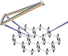
Scattering of X-rays from an antiferromagnetic ruthenate. The incident photons tuned to the Ru L3 absorption edge are focused on the sample, and interact with the Ru spins arranged in a two-dimensional honeycomb lattice. The scattered photons are dispersed with a newly-developed SiO2 diced spherical analyser (as illustrated by rainbow), and are collected with a CCD detector. The loss of X-ray energy (the change in “color”) is transferred to the system, which in turn excites the collective magnetic excitations. (Copyright: B. Keimer et al.)

An international team of scientists from the Max Planck Institute for Solid State Research, DESY, University of Stuttgart, Pohang University of Science and Technology POSTECH (South Korea) and the University of Tokyo (Japan) has determined the spectrum of collective magnetic excitations (“spin waves”) in a ruthenium-oxide antiferromagnet, which exhibits an unusually high magnetic ordering temperature. Such measurements are important because they yield insight into the magnetic interactions between spins inside the material, but they usually require large single crystals that are difficult to synthesize. By using the newly developed IRIXS spectrometer at PETRA III, the research team was now able to obtain a complete set of measurements on a microcrystal. The experiment demonstrates the capability of the IRIXS method - the probe of elementary excitations in a large class of magnetic materials.
Ruthenium and other 4d transition metal compounds play prominent roles in many areas of materials research ranging from electronics to catalysis. Furthermore, the systems can be used to investigate fundamental concepts in condensed matter physics, such as unconventional superconductivity, spin liquids, and solid-state analogues of the Higgs mode in particle physics. These collective quantum phenomena emerge as a result of a delicate interplay among the Coulomb interaction between the electrons, their interaction with the crystal lattice, and the intra-atomic spin-orbit coupling. However, basic questions about the mechanisms underlying these phenomena remain unanswered, because these key parameters are comparable in magnitude, and because sizable single crystals required for spectroscopic experiments are often difficult to synthesize.
To cope with this difficult task, scientists employed an experimental scheme called resonant inelastic X-ray scattering (RIXS). During the RIXS process, photons tuned to the absorption edge of a chemical element impinge on the sample and excite the system, which then undergoes a radiative decay to the final state. The energy of the photons emitted into a given direction is accurately measured, and the scattering intensity as a function of transferred energy and momentum is directly associated with the dispersion relations of elementary excitations of the system. Thanks to the significant energy and momentum carried by X-ray photons, charge, spin, and orbital excitations can be detected in a momentum-resolved and chemically selective manner. Crucially, the resonant condition greatly enhances the cross section of the RIXS process, enabling experiments on microcrystals and thin-film structures, which are not accessible to other spectroscopic probes such as inelastic neutron scattering.
Recent progress in the development of RIXS instrumentation in the soft and hard X-ray regimes has enabled comprehensive studies of magnetic compounds with 3d and 5d valence electrons, respectively. However, a RIXS instrument covering also the L-absorption edges of 4d transition metal compounds has not been available, since the absorption edges lie in the “intermediate” X-ray regime, where suitable optics have not been developed. A large and important class of magnetic materials was therefore not accessible so far. This has now changed after the construction of the IRIXS spectrometer at the dynamics Beamline P01 at PETRA III, which is equipped with a specifically designed energy analyzer for X-rays. The complementary capability to determine magnetic structures at Beamline P09 was also important for the success of this project.
The research team chose the antiferromagnet SrRu2O6, where Ru spins are arranged on a two-dimensional honeycomb lattice, to demonstrate the potential of the new instrument. The exceptionally high Néel temperature of this compound has attracted considerable recent attention, but spectroscopic experiments have thus far not been reported because only small single crystals (50 microns in diameter) could be synthesized. The IRIXS spectrometer enables measurements of the complete dispersion relation of spin waves, which allows to determine the interactions between the spins and their coupling to the crystal lattice. Their results were reported in Nature Materials.
The development of the IRIXS spectrometer was supported by the Advanced Grant “Com4Com” of the European Research Council (ERC).
Reference
Spin waves and spin-state transitions in a ruthenate high-temperature antiferromagnet; H. Suzuki, H. Gretarsson, H. Ishikawa, K. Ueda, Z. Yang, H. Liu, H. Kim, D. Kukusta, A. Yaresko, M. Minola, J. A. Sears, S. Francoual, H.-C. Wille, J. Nuss, H. Takagi, B. J. Kim, G. Khaliullin, H. Yavaş and B. Keimer; "Nature Materials" 2019; DOI:10.1038/s41563-019-0327-2