


")
")
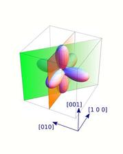
The three dimensional plot of the 3d orbital of Ni2+(from the last figure below (c)). (Credit:© MPI CPfS)

")
")
Orbital states are quantum mechanical constructions that describe the probability to find an electron in an atom, molecule or solid. We know from atomic physics that an s-orbital is spherical or that a p-orbital is dumbbell-shaped, but how do the complicated distributions of the electrons that contribute to chemical bonds in solids look like? Knowledge of these orbital states or electron distributions is the basis for our understanding of chemical bonds and related physical properties, which is a crucial step towards tailoring materials with specific characteristics. Here X-ray spectroscopy has contributed tremendously, however, the interpretation of the spectra is not easy and is often based on some assumptions for the analysis of the data. Hence it would be very important to have an experimental method that gives a direct image of the local electron density.
Such a method has been developed by the research teams of Prof. Liu Hao Tjeng (MPI-CPfS), Prof. Maurits Haverkort (University of Heidelberg) and Dr. Andrea Severing (University of Cologne) in a close collaboration with the PETRA III beamline P01. DESY scientist Hasan Yavas (now at SLAC (USA)), with the help of Martin Sundermann (DESY) and Hlynur Getarsson (DESY), built up an inelastic X-ray scattering end station that can be used for imaging local electron densities directly without the need for sophisticated mathematical analysis.
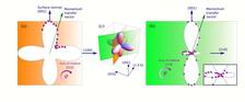
The experiments have shown at the example of Ni2+ in NiO, the textbook example for a strongly correlated material, that the angular dependence of the scattering intensity of the dipole forbidden 3s→3dtransition images directly the angular dependence of the 3A2 3d(x2-y2/3z2-r2) orbital of the nickel ion. This is based on the simple fact that the spherical3s orbital does not contribute to the angular distribution of the scattering intensity. The angular intensity of the scattering intensity is therefore a direct image of the more complex angular distribution of the local density of 3d electrons.
What sounds simple is experimentally not trivial because the transition s→d is dipole forbidden so that it cannot be reached with conventional methods like e.g. X-ray absorption. This transition, however, can be excited with photons when the energy transfer goes along with very large momentum transfers of the order of 10 Å-1. These large momentum transfers excite in addition to dipole so-called multipole transitions that have different selection rules so that even 3s→3d transitions have intensity. Such large momentum transfers can be reached is a non-resonant inelastic X-ray scattering experiment(NIXS), with hard X-rays of about 10 keV. The d-orbital can be imaged directly if the initial state is an s-orbital. This method is therefore called s-NIXS.
(from MPI-CPfS press release)
Reference:
Hasan Yavaş, Martin Sundermann, Kai Chen, Andrea Amorese, Andrea Severing, Hlynur Gretarsson, Maurits W. Haverkort & Liu Hao Tjeng, Direct imaging of orbitals in quantum materials, Nature Physics (2019)