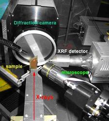
Figure 1:Experimental set-up adopted for combined µ-XRF/µ-XRD at Beamline L.
Roberto Terzano1, Matteo Spagnuolo1, Bart Vekemans2, Wout De Nolf2, Koen Janssens2, Gerald Falkenberg4, Saverio Fiore3, Pacifico Ruggiero1.
1Dipartimento di Biologia e Chimica Agro-forestale ed Ambientale, University of Bari, Bari, Italy.
2Department of Chemistry, University of Antwerp, Wilrijk, Belgium.
3Istituto di Metodologie per l’Analisi Ambientale (I.M.A.A.), C.N.R, Tito Scalo (PZ), Italy.
4HASYLAB at DESY, Hamburg, Germany.
Published as: R. Terzano et al., Environ. Sci. Technol., 41, 6762-6769.
It is commonly recognized that soil is the major sink for heavy metal (HM) contaminants released into the environment by human activities and that the mobility, bioavailability and toxicity of metals strongly depend on their solubility and therefore on their geochemical forms. Soils contaminated with HM can cause serious risks to human health, e.g. if vegetables cultivated on contaminated soils are consumed. The correct identification of HM chemical forms in soil is therefore of paramount relevance for a proper risk assessment and for the formulation of effective remediation strategies.
The major geochemical forms of Cr, Ni, Cu, Zn, Pb, and V in a soil from an industrial polluted site in the South of Italy were determined by means of synchrotron X-ray microanalytical techniques. On the basis of the geochemical forms identified, among the others, two major former industrial activities were tentatively ascribed as responsible of the observed major pollution: PVC and cement-asbestos productions.
In recent years, the development of new analytical methods exploiting high energy and high intensity synchrotron generated X-rays has provided soil scientists with new powerful tools for shedding light on HM speciation in soil [1-3]. In particular, given the large number of different phases in soils and their complex and heterogeneous distribution, the use of synchrotron X-ray microbeam techniques can resolve the metal bearing phases at the micrometer (or sub-micrometer) level thus allowing for a more selective and direct approach to HM speciation. These techniques have also the advantage of reducing analytical artefacts caused by extensive sample manipulations.
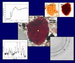
In this study, we used a combination of microanalytical techniques exploiting synchrotron generated X-rays as well as different bulk extraction methods to assess the geochemical forms and chemical behaviour of various HM pollutants [Cr, V, Pb, Ni, Cu, and Zn] in a soil from an industrial polluted site in the South of Italy. Micro X-ray Fluorescence (µ-XRF) was used to localize metals in soil thin sections and to find correlations among different elements. Simultaneously, micro X-ray diffraction (µ-XRD) patterns were collected to get information about the minerals HM were associated to. The simultaneous acquisition of µ-XRF and µ-XRD data was possible by collecting XRD patterns in transmission mode rather than in reflection mode [4]. In addition, µ-XANES spectroscopy point analyses were performed to determine the oxidation state of pollutants such as Cr and V and to better define the association of various other metals with minerals, by comparison with selected mineral standards. The information revealed by these synchrotron radiation based analyses allowed for HM speciation at the microscopic level.
All the results emerging from both the “microscopic” (X-ray based) and the “macroscopic” (bulk extraction based) investigations were combined and employed to identify the possible former sources of metal pollution and to predict the fate of these elements in soil.
The experimental set-up adopted for the combined microanalytical investigation at Beamline L (HASYLAB) is reported in Figure 1.
|
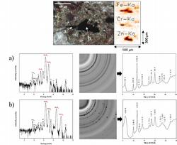
Several micro-areas on different soil thin sections were analysed by means of combined synchrotron µ-XRF/µ-XRD (Figure 2). |
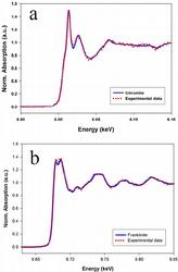
In addition, for selected points of interest, metal speciation was more clearly assessed by µ-XANES (Figure 3). |
|
Figure 2: Micrograph with the corresponding µ-XRF distribution-maps (for Fe, Cr, and Zn) of a microscopic area on a soil thin section analysed by combined µ-XRF/µ-XRD. Darker pixels correspond to relative higher concentrations of the element. µ-XRF spectra and µ-XRD images (also converted into 1D diffraction patterns) were collected in points a and b (indicated by the arrows in the micrograph). The d-spacing values corresponding to the diffraction peaks are also shown in the 1D diffractogram. The reported d-spacings are characteristic of spinel-type minerals: a) chromite and/or magnetite, b) franklinite and/or magnetite. |
|
Using this approach, various mineral phases were microscopically identified for the different metals, as reported in Table 1.
Major elements (%) |
Trace elements (µg/g) |
Microscopically identified phases |
||
|---|---|---|---|---|
SiO2† |
40.1 % |
Crtot* |
352±37 |
Chromite, Zincochromite, sorbed on hematite |
TiO2† |
0.6 % |
Cr(VI)alk* |
5.1±0.9 |
Crocoite |
Al2O3† |
9.8 % |
Cr(VI)ac* |
12±2 |
|
Fe2O3† |
16.4 % |
Zn* |
1196±53 |
Franklinite, Zincochromite, sorbed on hematite |
MnO† |
0.3 % |
Ni* |
394±17 |
Trevorite, sorbed on hematite |
MgO† |
1.4 % |
Cu* |
449±8 |
Cuprospinel, Volborthite, sorbed on hematite |
CaO† |
18.1 % |
Pb* |
1080±150 |
Minium, Lanarkite, Crocoite |
Na2O† |
0.6 % |
V* |
1688±24 |
Volborthite, V(V) sorbed on hematite |
K2O† |
1.1 % |
Ba* |
445±12 |
Barite |
L.O.I. |
11.6 % |
|
|
|
| † Determined by XRF | alk: alkaline extraction |
| *Determined by ICP-AES | ac: acidic extraction |
Cr, Ni, Zn and Cu were found in spinel-type geochemical forms (chromite, trevorite, franklinite, zincochromite, cuprospinel) and often in association with magnetite and hematite. V was mainly present as V (V) associated to Fe-oxides or in the form of volborthite [Cu3(OH)2V2O7•2H2O]. Pb was speciated as minium (Pb3O4), lanarkite [Pb2O(SO4)] and, in association with Cr(VI), as crocoite (PbCrO4).
The main limit of such microanalytical method is represented by the difficulty to reliably extrapolate the obtained results to a higher length scale. Therefore, to analyze larger amounts of soil samples, various extraction procedures have been also adopted, including single or sequential extraction steps. Most of the results obtained at the microscopic level have been confirmed by the extraction procedures. However, batch extractions failed to correctly identify some HM chemical species (in particular for Pb). Therefore, for the analysed soil samples, an extrapolation of the results obtained by using microspectroscopic techniques to the macroscopic level seems to be applicable even if most of the well-known limitations of the extraction procedures are confirmed [5].
From all the data presented in this research, despite a high total concentration, most of the HM appear to be speciated in rather insoluble geochemical forms. Therefore, the environmental danger connected to their presence in the investigated soil is estimated to be much lower than expected. However, particular attention should be still paid to Zn, Cu, V, and Pb that show non negligible mobilizable fractions, as evidenced by EDTA extractions.
Finally, the detailed HM speciation determined by microspectroscopic techniques has been compared with the geochemical characteristics of the site under investigation and the possible sources of anthropogenic impact have been determined.
In particular, two major former industrial activities were tentatively ascribed as responsible of the observed major pollution: PVC and cement-asbestos productions.
Acknowledgements
This research was financed by the MIUR (COFIN 2005) project “Innovative chemical, physical, and biological methods to characterize and remediate soils polluted by heavy metals (MICROS)” and by the IMAA-CNR project “Elemental Mobility Geochemical Map in Basilicata-Italy”. Synchrotron experiments at HASYLAB were financially supported by the European Community-Research Infrastructure Action under the FP6 “Structuring the European Research Area” Program (Integrating Activity on Synchrotron and Free Electron Laser Science; project: contract RII3-CT-2004-506008).
|
References |
Contact information |
|
[1] Bernaus, A.; Gaona, X.; Esbrì, J.M.; Higueras, P.; Falkenberg, G.; Valiente, M. Environ. Sci. Technol. 2006, 40, 4090-4095. |
Roberto Terzano (corresponding author) |
| Further Information |


