


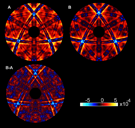
Figure 1: Angular fine structure of white x-ray absorption recorded for a GaP crystal for two opposite crystal orientations (A and B). The bottom pattern is the difference image. These patterns correspond to distorted real-space views (“shadowgrams”) of the GaP crystal. The intensity scale is normalized to the slowly varying background of the x-ray absorption.
P. Korecki1, M. Tolkiehn2, D.V. Novikov2, G. Materlik3, M. Szymonski1
1M. Smoluchowski Institute of Physics, Jagiellonian University, Krakow, Poland
2Hamburger Synchrotronstrahlungslabor HASYLAB at DESY, Hamburg, Germany
3Diamond Light Source Limited, Chilton, Oxfordshire, United Kingdom
Published as: P. Korecki et al., Physical Review Letters 96, 035502 (2006)
Conventional x-ray crystallographic methods, which are used for almost 100 years, detect x-rays diffracted by the crystal planes. In a recent paper it was demonstrated that the atomic structure can be imaged directly from real-space projections sensed by absorbing atoms inside a crystal. The use of white x-rays allowed neglecting the diffraction effects and treating the x-ray beam as a searchlight which directly produces x-ray projections of the main atomic planes in the crystal. The recorded x-ray patterns were processed in a way similar to the tomography technique which is used e.g. for 3D visualization of macroscopic objects in medical imaging.
Most of x-ray methods for crystal structure investigations are based on diffraction phenomena and sample the information in the reciprocal space using an external far-field detector. In principle, transformation from reciprocal to real-space can be made using a Fourier transformation. However, such a direct back transform is often hindered by the lack of phase information in the recorded intensity data so that the inversion algorithms become ambiguous.
An alternative approach to atomic resolution imaging involves measuring the x-ray wave field intensity directly inside a crystal, at atomic sites. Such a measurement is performed in the x-ray standing waves technique [1] or in atomic resolution x-ray holography [2-4]. The absorption cross-section in these methods is modulated by x-ray diffraction, which results in an angular dependent absorption fine structure measured through the secondary yield coming from specific sorts of atoms. Probing of the interference of the beams inside the sample can give access to the relative phase of the scattered waves in a way similar to holography. This angular fine structure can be inverted to real space by using Fourier or holographic reconstruction. These methods utilize monochromatic x-ray radiation.
In our works [5,6] it was demonstrated that the patterns of the angular fine structure in x-ray absorption recorded using white x-rays have a simple real-space interpretation. For a white x-ray beam, the variations of the wave field, formed by interference of the incident beam with the waves scattered on single atoms, cancel out by energy integration for all directions, except for the nearly forward scattering components, coinciding with the incident beam.
The experiments were carried out at the bending magnet beamline CEMO at HASYLAB. Fig.1 shows the angular dependence of white x-ray absorption measured for a GaP crystal via photoelectron current. Each point in the patterns correspond to a particular orientation of the sample relative to the incident beam. The presented absorption patterns show distorted projections of main atomic planes in the crystal. This small distortion results from remnant high-order diffraction effects caused by a non-uniform x-ray energy spectrum.
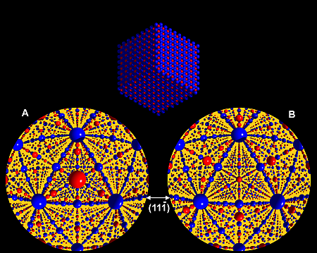
For comparison, inside views of the GaP crystal observed from a position of a single Ga atom are presented in Fig.2 Note the similarities between the experimental patterns and fish-eye inside views. The low-intensity points in the recorded patterns (shown in cold colors) correspond to high density directions in the fish-eye images, whereas the high intensity points (hot colors) to empty spaces in the crystal. The positions of Ga atoms are the same and the positions of P planes are different for both crystal orientations. In particular, P planes lie outward and inward of the triangle set by the {111} planes in fish-eye images A and B. This difference is directly observable in the experimental patterns as well.
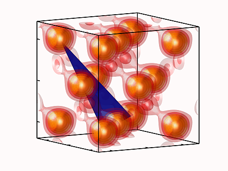
The reconstruction of the object from its projection is the domain of tomography. Thus, the measured patterns were processed using a newly developed tomographic algorithm [6] and allowed for a visualization of the unit cell of GaP with spatial resolution better than 0.1 nm as shown in Fig.3.
In future the presented approach could be applied for imaging of dopants atom positions and for imaging of thin films, interfaces and buried layers.
| References |
|
[1] J. Zegenhagen, Surf. Sci. Rep.18, 199 (1993) |
| Contact information |
|
P.Korecki |
| Further Information |