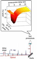
FIG. 1: Transient X-ray induced optical reflectivity (ΔR/R) measurement - schematic overview:
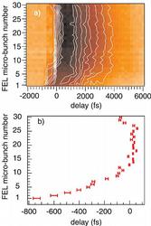
Extreme ultraviolet FEL pulses (39.5 eV, < 50 fs, < 16µJ) impinge onto a crystalline GaAs(100) surface and generate photoexcited carriers. The transient changes of the dielectric function are probed by visible laser pulses (800 nm or 400 nm, 120 fs, < 10 nJ ) reflected from the GaAs surface at 53° as a function of their temporal delay relative to the FEL radiation pulse. The visible laser operates at twice the repetition rate (1 MHz) of the FEL (500 kHz) to measure the pumped and unpumped surface as a reference. Experimental reflectivity data for a bunchtrain of 30 microbunches is shown.
C. Gahl,1,3 A. Azima,5 M. Beye,2 M. Deppe,2 K. Döbrich,1 U. Hasslinger,2 F. Hennies,2,4 A. Melnikov,1 M Nagasono,2 A. Pietzsch,2 M. Wolf,1 W. Wurth,2 and A. Föhlisch2 *
1 Fachbereich Physik, Freie Universität Berlin, Berlin, Arnimallee 14, 14195 Berlin, Germany
2 Institut für Experimentalphysik, Universität Hamburg, Luruper Chaussee 149, 22761 Hamburg, Germany
3 Max-Born-Institut für nichtlineare Optik und Kurzzeitphysik, Max-Born-Str. 2A, 12489 Berlin, Germany
4 MAX-lab, Lund Universitet, Ole Römers väg 1, Box 118, 221 00 Lund, Sweden
5 HASYLAB/DESY, Notkestr. 85, 22607 Hamburg, Germany
Published as: C. Gahl et al., Nature Photonics 2, 165-169 (2008)
For a fundamental understanding of ultra fast dynamics in chemistry, biology and materials science it has been a longstanding dream to record a molecular movie, where both the atomic trajectories and the chemical state of every atom in matter is followed in real time with time resolved pump/probe spectroscopy. X-ray free-electron lasers (FEL) provide this perspective as they deliver brilliant femtosecond X-ray pulses spanning a wide photon energy range. To cross-correlate and synchronize the FEL with separate optical lasers we exploit the peak brilliance of the free-electron laser at Hamburg (FLASH) and establish X-ray pulse induced transient changes of the optical reflectivity in GaAs as a powerful tool for X-ray/optical cross-correlation. This constitutes a breakthrough en route to a molecular movie and – equally important – opens the novel field of femtosecond X-ray induced dynamics, only accessible with high brilliance X-ray free-electron lasers.
Introduction
Free-electron lasers like FLASH1,2 at DESY excel in producing femtosecond X-ray bursts of variable photon energy at maximum peak brilliance and coherence using the principle of self amplification of spontaneous emission (SASE).3,4,5,6,7 Combined with optical lasers X-ray spectroscopic and diffractive methods allow to study ultra fast photo induced dynamical processes from low energy IR activated dynamics to excitations in the optical and UV spectral range. In particular the selectivity of X-ray spectroscopy enables to monitor the temporal evolution of few selected active sites without the background of a large number of surrounding atoms from a passive matrix, as resonant X-ray excitation yields atom specific and chemical state selective information8,9,10,11,12. Thus disperse systems, highly relevant in biology and chemistry, e.g. in heterogeneous catalysis and colloidal systems, become accessible. Furthermore, the high peak brilliance provides sufficient X-ray pulse energy to study X-ray induced dynamics for the first time. This paves a new route to radiation chemistry and to study, for example, the radiation induced processes at interstellar dust or the chemistry at particles in the upper atmosphere.13,14 A crucial aspect for these investigations is to measure the synchronization between the X – ray pulses with optical or infrared pulses from an external laser source with femtosecond accuracy at the interaction point within an experimental set up. An important first step has been achieved by measuring the relative arrival time of the electron bunches within the linear accelerator structure through electro optical sampling (EOS)21. However, since the overall dimensions of FEL sources require to guide radiation pulses over approx. 100 m via various optical elements in separate X-ray and optical beamlines, we need to measure the precise time delay and spatial overlap directly at the interaction point of the experimental set-up. Such a cross-correlation measurement would allow eliminating the temporal jitter between the optical and X-ray laser pulses. One approach is to measure optical laser induced side band generation in vacuum ultraviolet photoemission from noble gases.17,15 This can only be done using dedicated electron spectrometers under high vacuum conditions14 and is limited by space charge effects. Second harmonic and sum frequency generation, routinely used to correlate optical pulses from different lasers16, suffer – when applied to X – rays – from low cross sections.
In this work, we have now established ultra fast transient changes of the optical reflectivity in GaAs induced by femtosecond X-ray excitation as an ideal tool for X – ray/optical cross correlation, which can be applied to any sample environment spanning the energy range of present and future FEL X–ray sources.
Strategy of the Experiment
Using the Hamburg inelastic X – ray scattering station on the plane grating monochromator beamline PG2 at FLASH17 we reflected visible femtosecond laser pulses off a clean, undoped GaAs(100) surface and measured the temporal change of the optical reflectivity as it is modified through brilliant extreme ultraviolet radiation from FLASH. As shown in Fig. 1, the induced change of optical reflectivity was probed at an angle of incidence of 53° by delayed optical pulses at 800 nm or optionally 400 nm with a duration of 120 - 150 fs (fwhm) delivered from an optical parametric amplifier system with 1 MHz repetition rate, electronically synchronized to the electron accelerator18. The optical pulse energies were detected in a reference path and after reflection with two fast photodiodes, allowing for transient reflectivity measurements pulse by pulse using a 2 GHz analog to digital converter (Acqiris) embedded into the distributed object oriented control system (DOOCS)19 of FLASH. The FEL was operated at a macro bunch repetition rate of 5 Hz consisting of bunch-trains of 30 micro-bunches with 2µs separation (500 kHz). Operating the optical laser at twice the FEL repetition rate within each pulse train of 30 radiation bursts results in alternating measurements of the reflectivity with and without the X-ray pump pulse. The FLASH pulses at 39.5 ± 0.5 eV with a duration of < 50 fs impinge on the GaAs(100) crystal at 41.5° incidence angle with pulse energies up to 16µJ. Thus at a spot size of (395 ± 23)µm x (274 ± 14)µm 20 the fluence stays well below the optical damage threshold of the GaAs surface (50 mJ/cm2 for 30 fs pulse length at 800 nm)21.
Timescales of X-ray induced dynamics and their physical origin
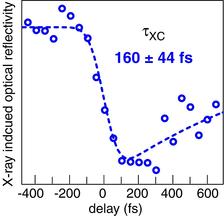
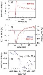
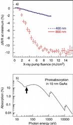
In Fig. 2 we present the FEL X-ray pulse induced transient optical reflectivity changes ΔR/R on time scales from femtoseconds to many hundreds of picoseconds, where from panel 2 a) to 2 c) different temporal delay ranges are presented. The intense X-ray excitation leads to an ultra fast drop in optical reflectivity, which recovers within a few picoseconds. Depending on the X-ray fluence and probe wavelength, ΔR/R can even overshoot to positive values before the system approaches equilibrium on the time scale of more than 100 ps as shown for 800 nm probe. In the following, we focus on the femtosecond time scale of the ΔR/R transients, which is of key relevance for cross-correlation measurements of X – ray and optical radiation pulses. Fig. 2 b) shows that the rapid X-ray pulse induced drop in optical reflectivity occurs on the same time scale for both 800 nm and 400 nm probe wavelength, respectively. The time scale can be deduced from a measurement shown in Fig. 2 c), where we corrected for the major sources of temporal jitter within the accelerator (typically 0.25 ps RMS 14) using electro optical sampling22 to determine the arrival time of selected electron bunches relative to the optical laser. When we fit the reflectivity transient with an exponentially decaying response function convoluted with a Gaussian we obtain for the latter a full width at half maximum of 160 ± 44 fs. Thus the intrinsic time constant of the X – ray pulse induced initial drop in optical reflectivity is small compared to the cross-correlation width making it suitable for cross-correlation measurements of FEL X-ray and optical laser pulses. To understand the physical origin of the ultra fast reflectivity drop we turn to the X-ray pump fluence and X-ray pump energy dependence as summarized in Fig. 3). At 800 nm probe wavelength, where in equilibrium only optical transitions across the direct band gap close to the Γ point are probed23, the minimum of the ΔR/R transient scales linearly with the fluence up to 6 mJ/cm2 and then stays almost constant. At 400 nm probe wavelength a smaller amplitude of the ΔR/R transient minimum is found and optical transitions at many k-values between the L – and Γ – points in the band structure are possible22. The absorption of the FEL radiation is governed by the atomic photoionization cross sections (Ga 3d: 3.6 Mbarn, Ga 4s: 0.17 Mbarn, As 4p: 0.3 Mbarn, As 4s: 0.21 Mbarn at 40.8 eV)24,25 leading preferentially to Ga 3d vacancies. Within 10 nm of the GaAs sample 10% of the incident photons are absorbed26 (Fig. 3b), staying below the damage threshold of the GaAs surface (50 mJ/cm2)20. Thus an excitation fluence of 10 mJ/cm2 (equivalent to 1.6•1015 photons at hv=39.5 eV) creates within the 10 nm thick surface layer a Ga 3d excitation density of approx. 1.6•1020 cm-3. Within few femtoseconds Auger decay and autoionization converts the initial inner shell excitation27 into twice the number of valence excitations (~ 3•1020 cm-3) and finally equilibration to the mean energy of electron-hole pair creation in GaAs of 4.2 eV28 produces an electron-hole pair density of 1.5•1021 cm-3. With optical laser excitation at and above the optical damage-threshold photo-generated free carriers exceeding 1020 cm-3 lead to similar ΔR/R transients as we find for X-ray excitation below the damage threshold. There the free carrier absorption changes the dielectric function as described by the Drude model. Beyond that, screening of ionic potentials and electron many-body effects are important as they modify the band structure22,29. Thus a full theoretical model of the X-ray pulse induced optical transient reflectivity will consider the response of the dielectric function to the distortion of the valence electronic structure through photoionization and ultra fast Auger decay of inner shell vacancies, electronic screening and electron-electron scattering as well as the structural changes to the crystal lattice.
An X-ray/optical cross-correlation monitor for FLASH
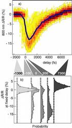
As a first application we monitor the temporal overlap between optical and X – ray laser pulses over an extended period of time (Fig. 4). Due to the temporal jitter within the accelerator, we observe at a nominally fixed pump-probe delay a characteristic distribution of the optical reflectivity signal (Fig. 4). Characteristic differences in the ΔR/R intensity distributions are found for different delay positions in the femtosecond and picosecond range. In particular, around time zero the temporal jitter is translated into strong variations of the optical reflectivity. The reflectivity signal changes already significantly for a delay difference of 100 fs or 200 fs allowing for an online monitoring of pulse-to-pulse jitter. At larger temporal delay further variations of the ΔR/R intensity distribution function of optical reflectivity are found. The presented technique has been extended to single shot X – ray pulse diagnostics by imaging the reflected optical pulses onto a spatially resolving detector mapping arrival time onto a spatial coordinate30. In such a crossed beam experiment the temporal relation between X – ray and optical pulses is measured fully independent from any other temporal measurement. Finally, it might be possible to achieve full temporal and energetic characterisation of every radiation pulse by using an X-ray diffraction grating or crystal, where zero order light is delivered to the user experiment and parasitic to user operation, the first order spectral image is used to project energy and time information onto orthogonal axes.
The temporal fine-structure within FLASH bunch trains
We can now also apply our X-ray/optical cross correlator to investigate temporal characteristics of the FLASH X-ray pulse trains. In Fig. 5 a) the transient optical reflectivity measurement is shown for FLASH operating with 30 micro bunches thus emitting 30 subsequent FEL femtosecond X – ray pulses separated by 2 µs each. As shown in Fig. 5 b) we observe a significant systematic drift of the delay by almost 1 ps over the first part of the pulse train stabilizing after ~10 pulses. Since this delay drift is caused by the electronic feedback systems of the accelerator structure and the coupling of subsequent electron bunches, it also varies with the operation condition of the accelerator. Thus its characterization and optimization are especially important for pump-probe experiments on a femtosecond timescale. Knowledge about the precise timing allows using drifts for a complete delay scan within a single bunch train or to alternatively correct the arrival time of all micro bunches. The investigation of these systematic timing drifts within the X-ray pulse trains could not be done before, as methods like electro optical sampling have been too slow in data acquisition for the 500 kHz repetition rate of FLASH.
Relevance for future X-ray FEL sources
An important aspect is how X-ray induced transient optical reflectivity can be used for the extended energy range of present and future FEL sources. Based on the physical mechanism explained previously, we present in Fig. 3b) the photoabsorption in 10 nm GaAs as the fraction of photons absorbed. As the photoabsorption decreases less than inversely with photon energy, the reduced absorption probability is compensated by the higher photon energy, as the density of the excited carrier distribution responsible for the changes in optical reflectivity also depends on the absorbed energy. Taking our measurement at 39.5 eV photon energy as the reference, we expect comparable or higher ΔR/R signal strength up to 6.5 keV and at most a reduction by a factor of two up to 15 keV. Thus X–ray/optical cross correlation by means of transient optical reflectivity measurements should allow to easily monitor the temporal relation between FEL X–ray pulses and optical laser pulses at the interaction point within user experiments, suitable for a large variety of experimental conditions.
Summary
The technique of X – ray induced transient optical reflectivity on a GaAs surface has been established as a powerful tool for cross correlation between femtosecond optical and X – ray pulses, spanning the energy range of present and future X-ray free-electron laser sources. Furthermore, X-ray induced non-equilibrium dynamics opens a new field of time resolved studies of matter which is highly relevant for X-ray induced chemistry in biological systems, solids and interfaces e.g. present in atmospheric and interstellar dust. Thus our findings pave the way towards time resolved structural dynamics in chemistry, biology and materials science.
We dedicate this manuscript to our suddenly deceased co-worker Kai Starke, who’s enthusiasm and competence was essential to initiate this work. We thank Christian Kumpf (University of Würzburg) for providing the GaAs samples. This work was supported by the German Ministry of Education and Research (BMBF) through Grants No. 05 KS4GU1/8 and No. 05 KS4GU1/9 and the Helmholtz Joint Research Centre “Physics with coherent radiation sources”.
|
References |
|
1 V. Ayvazyan et al., First operation of a Free-Electron Laser generating GW power radiation at 32 nm wavelength, Eur. Phys. J. D 37, 297-303 (2006). 2 W. Ackermann et al. Operation of a free-electron laser from the extreme ultraviolet to the water window, Nature Photonics 1, 336-342 (2007). 3 J. M. J. Madey, Stimulated emission of bremsstrahlung in a periodic magnetic field, J. Appl. Phys. 42, 1906-1913 (1971). 4 D. A. G. Deacon, L. R. Elias, J. M. J. Madey, G. J. Ramian, H. A. Schwettman, and T. I. Smith, First Operation of a Free-Electron Laser, Phys. Rev. Lett. 38, 892-894 (1977). 5 A. M. Kondratenko and E. L. Saldin, Generation of coherent radiation by a relativistic electron beam in an undulator, Soviet Phys. Doklady 24, 986-988 (1979). 6 A. M. Kondratenko and E. L. Saldin, Generation of coherent radiation by a relativistic electron beam in an ondulator,Part. Acc. 10, 207-216 (1980). 7 R. Bonifacio, C. Pellegrini, L. Narducci, Collective instabilities and high-gain regime in a free electron laser, Opt. Commun. 50, 373-378 (1984). 8 A. Föhlisch, N. Wassdahl, J. Hasselström, O. Karis, D. Menzel, N. Mårtensson, and A. Nilsson, Beyond the Chemical Shift: Vibrationally Resolved Core-Level Photoelectron Spectra of Adsorbed CO, Phys. Rev. Lett. 81, 1730-1733 (1998). 9 A. Föhlisch, M. Nyberg, J. Hasselström, O. Karis, L.G.M. Pettersson, and A. Nilsson, How Carbon Monoxide Adsorbs in Different Sites, Phys. Rev. Lett., 85, 3309-3312 (2000). 10 J. T. Lau, A. Föhlisch, R. Nietubyc, M. Reif, and W. Wurth, Size-Dependent Magnetism of Deposited Small Iron Clusters Studied by X-Ray Magnetic Circular Dichroism, Phys. Rev. Lett., 89, 0572011(1-4) (2002). 11 A. Föhlisch, P. Feulner, F. Hennies, A. Fink, D. Menzel, D. Sanchez-Portal, P. M. Echenique, and W. Wurth, Direct observation of electron dynamics in the attosecond domain, Nature 436, 373-376 (2005). 12 A. Pietzsch, et al,Towards time resolved core level photoelectron spectroscopy with femtosecond X-ray free-electron lasers,New J. Phys. 10 033004 (2008). 13 D. C. Lis, G. A. Blake, E. Herbst, Astrochemistry: Recent Successes and Current Challenges, International Astronomical Union, Cambridge University Press (2006). 14 J. M. Greenberg, J. S. Gillette, G. M. Muñoz Caro, T. B. Mahajan, R. N. Zare, A. Li, W. A. Schutte, M. de Groot, and C. Mendoza-Gómez, Ultraviolet Photoprocessing of Interstellar Dust Mantles as a Source of Polycyclic Aromatic Hydrocarbons and Other Conjugated Molecules, The Astrophysical Journal, 531, L71–L73 (2000). 15 S. Cunovic et al. Time-to-space mapping in an gas medium for the temporal characterization of vacuum-ultraviolet pulses, Appl. Phys. Lett. 90, 121112(1-3) (2007). 16 J.-C. Diels and W. Rudolph, Ultrashort Laser Pulse Phenomena, Academic Press Inc., U.S. 2nd revised edition (2006). 17 M. Wellhöfer, M. Martins, W. Wurth, A. A. Sorokin and M. Richter, Performance of the monochromator beamline at FLASH, J. Opt. A: Pure Appl. Opt. 9, 749-756 (2007). 18 P. Radcliffe et al., Single–shot characterization of independent femtosecond extreme ultraviolet free electron and infrared laser pulses, Appl. Phys. Lett. 90, 131108(1-3) (2007). 20 S. Düsterer et al. Spectroscopic characterization of vacuum ultraviolet free electron laser pulses, Opt. Lett. 31, 1750-1752 (2006). 21 A. Cavalleri, et al., Ultra fast x-ray measurement of laser heating in semiconductors: Parameters determining the melting threshold, Phys. Rev. B 63, 193306(1-4) (2001). 22 A. L. Cavalieri et al., Clocking Femtosecond X-Rays, Phys. Rev. Lett. 94, 114801(1-4) (2005). 23 J. P. Callan, A. M.-T. Kim, L. Huang, E. Mazur, Ultra fast electron and lattice dynamics in semiconductors at high excited carrier densities Chemical Physics 251, 167-179 (2000). 24 J.-J. Yeh and I. Lindau, Atomic Subshell Photo ionization Cross Sections and Asymmetry Parameters: 1 < Z < 103, At. Data Nucl. Data Tables 32, 1 (1985). 25 J.-J. Yeh, Atomic Calculations of Photo ionization Cross Sections and Asymmetry Parameters, Gordon and Breach, Langhorne, PA (1993). 26 B. L. Henke, E. M. Gullikson, and J. C. Davis, X – ray Interactions: Photoabsorption, Scattering, Transmission, and Reflection at E = 50 – 30000eV, Z=1-92, At. Data Nucl. Data Tables 54, 181-342 (1993). 27 M. O. Krause and J. H. Oliver, Natural widths of atomic K and L levels, K alpha X-ray lines and several KLL Auger lines, J. Phys. Chem. Ref. Data 8, 329-338 (1979). 28 M. Krumrey, E. Tegeler, J. Barth, M. Krisch, F. Schäfers, and R. Wolf, Schottky type photodiodes as detectors in the VUV and soft X-ray range, Applied Optics 27, 4336-4341 (1988). 29 L. Huang, J. P. Callan, E. N. Glezer, and E. Mazur, GaAs under intense ultra fast excitation: Response of the dielectric function, Phys. Rev. Lett. 80, 185-188 (1998). 30 T. Maltezopoulos et al. Single shot timing measurement of extreme-ultraviolet free-electron laser pulses, New J. Phys. 10, 033026 (2008). |
|
Contact information |
|
PD PhD Alexander Föhlisch |
| Further Information |


