


")
")
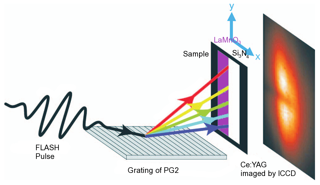
Figure 1: Experimental set-up at FLASH: The pulses of FLASH are dispersed by a grating-monochromator. The sample intercepts the beam. One half of the beam is transmitted through the Si3N4-membrane, while the half on the side of the LaMnO3-film is partly absorbed.
D.P. Bernstein1, Y. Acremann1, A. Scherz1, M. Burkhardt1, J. Stöhr1, M. Beye2, W.F. Schlotter2, T. Beeck2, F. Sorgenfrei2, A. Pietzsch2, W. Wurth2, and A. Föhlisch2 (now at 3, 4 )
1. SLAC National Accelerator Laboratory, 2575 Sand Hill Road, Menlo Park, California 94025, USA
2. Institut für Experimentalphysik and Centre for Free Elecotron Laser Science (CFEL) Universität Hamburg , Luruper Chaussee 149, 22761 Hamburg, Germany
3. Institute for Methods and Instrumentation in Synchrotron Radiation Research G-I2, Helmholtz-Zentrum Berlin für Materialien und Energie, Albert-Einstein-Str. 15, 12489 Berlin, Germany
4. Institut für Physik und Astronomie, Universität Potsdam, Karl-Liebknecht-Strasse 24-25, 14476 Potsdam, Germany
Published as: “Near edge X-ray absorption fine structure spectroscopy with X-ray free-electron lasers”, Appl. Phys. Lett. 95, 134102 (2009).
A new method to perform X-ray absorption spectroscopy experiments at a free-electron laser has been developed. Instead of selecting a narrow bandwidth of the incident beam with a grating-monochromator, a dispersive set-up is used. The incident pink radiation from the SASE-FEL is dispersed by the grating and collected by an area detector. A special sample-preparation method has been used in order to measure the intensity and energy distribution of the incident and absorbed beam simultaneously. This method can be improved in the future to perform pump-probe experiments with XAS as the probe with fs-temporal resolution at an FEL.
Near edge X-ray absorption spectroscopy (NEXAFS or XANES) is uniquely powerful [1-3] to learn about the electronic structure properties of matter, which determines materials properties like magnetism. With femtosecond X-ray pulses, transient states and ultra-fast dynamics in matter are accessible and have been explored so far at femtosecond slicing facilities at synchrotron radiation facilities. Turning to the brilliant femtosecond X-ray pulses from Free-Electron Laser Sources we now want to expand femtosecond time resolved electronic structure studies further and explore how brilliant X-ray pulses modify electronic properties in matter.
To this end, we adapted the way NEXAFS is measured to the requirements imposed by the nature of SASE-FEL radiation. The main point is that we do not scan the monochromator, but use it in dispersive geometry, where the spectrum is then measured by an area detector, where the position on the detector corresponds to one specific energy. For our experiment we chose a thin film of LaMnO3 (75 nm) deposited by pulsed laser deposition on a thin Si3N4 membrane (100nm) in such a way that the LaMnO3 film covered just one half of the membrane [4]. The other half was left intentionally blank in order to determine the spectral distribution of the incident beam as a reference.
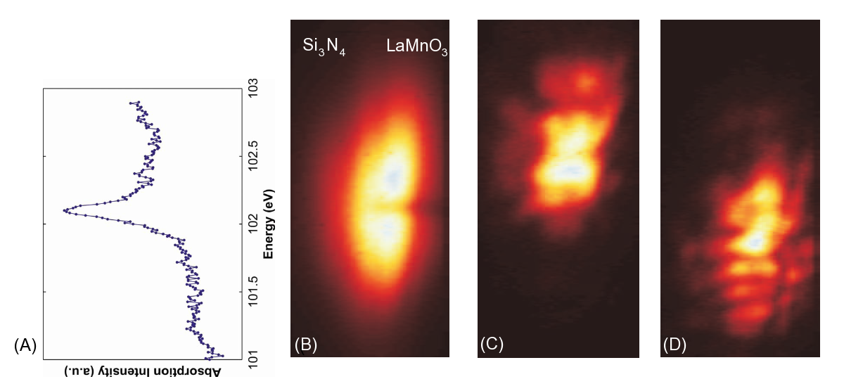
The beauty of this system is, that the N4,5 edge of the La3+ ions in LaMnO3 exhibits an atomically sharp 3D1 absorption resonance at a photon energy of 102.17 eV, where the Si3N4 membrane is highly transparent. The radiation-fan from the grating at the PG2 beamline at FLASH [5] is passing through the sample, where one-half of the fan is absorbed by the LaMnO3 film and the membrane and the other passed through the uncovered membrane. The half that is transmitted through the membrane is used for determining the intensity and spectral distribution of the incident flashes, the other half absorbs the radiation based on its energy. Since energy and intensity are measured simultaneously, it is possible to normalise each individual spectrum, which reflects only a small part of the total absorption spectrum.
These measurements have brought us a good step towards femtosecond time resolved NEXAFS for materials science and high field induced studies in solids. Future improvements will aim to bring the interaction region just between the X-ray source and the monochromator grating in order to ensure the shortest possible and eventually the most brilliant X-ray pulses to interact with the sample.
| References | ||||||||||
|
| Contact information |
|
Alexander Föhlisch |
| Further Information |