


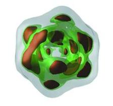
Particle reconstruction results: 3D rendering of inner surfaces. (Credit: DESY)
")
")
An international team of researchers, led by scientists from DESY and Kurchatov Institute (Moscow, Russia), has presented a workflow from single-particle imaging (SPI) data, measured at X-ray free-electron lasers (XFELs), to a three-dimensional reconstruction of the structure. They published their findings in the journal of the International Union of Crystallography (IUCrJ).
Single-particle imaging by using hard XFELs is a method to get the structural information of relevant biological systems, like viruses. This promising method was proposed about 20 years ago and is still under development. The challenge is to gain the structural information from a huge amount of data sets. They are created during the experiment, when a jet of randomly oriented and free movable biological particles is hit by a XFEL pulse. In contrast to cryo-electron microscopy, the single particles do not need to be frozen at liquid nitrogen temperatures to determine their structure. However, to achieve a high-resolution or even atomic resolution structure determination of such biological particles using XFELs is more demanding than expected.
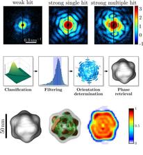
To improve this method, the international team used data measured at the Atomic Molecular Optics (AMO) instrument at the Linac Coherent Light Source LCLS at SLAC in Stanford (USA). This was part of the SPI initiative headed by LCLS. As a sample the PR772 bacterio-phage was chosen, with a diameter of about 70 nm. Their workflow consisted of several steps, which enabled the analysis of large SPI data sets, including single-hit diffraction data classification, refined filtering of the classified data, reconstruction of three-dimensional scattered intensity from the experimental diffraction patterns by orientation determination and a final three-dimensional reconstruction of the virus electron density without symmetry constraints.
As a result of these analysis steps, features of the virus could be obtained with an resolution better than 10 nm, which could not be reached so far. Moreover the three-dimensional reconstruction shows, that the virus was compressed in one direction, which breaks the ideal icosahedral symmetry.
Original Publication: “Single-particle imaging without symmetry constraints at an X-ray free-electron laser“, M. Rose, S. Bobkov, K. Ayyer, R. P. Kurta, D. Dzhigaev, Y. Yong Kim, A. J. Morgan, C. Hong Yoon, D. Westphal, J. Bielecki, J. A. Sellberg, G. Williams, F. R.N.C. Maia, O. M. Yefanov, V. Ilyin, A. P. Mancuso, H. N. Chapman, B. G. Hogue, A. Aquila, A. Barty and I. A. Vartanyants, IUCrJ, Vol. 5, Part 6 (2018), DOI: 10.1107/S205225251801120X