


")
")
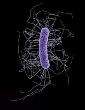
Electron microscope image of a Clostridium difficile bacterium. (Credit: Jennifer Hulsey/CDC)
Research at DESY's X-ray light source PETRA III reveals how certain viruses called bacteriophages kill the potentially life threatening diarrhoea bacterium Clostridium difficile. The study by scientists of the European Molecular Biology Laboratory (EMBL) in Hamburg shows how enzymes of these viruses are triggered and released to degrade the cell walls of the bacteria. The work opens up new opportunities for developing effective bacteriophage therapies, as the researchers led by EMBL Group Leader Rob Meijers write in the scientific journal PLoS Pathogens.
“In the light of increased antibiotic resistance, bacteriophages and their enzymes could provide a good alternative,” says Meijers. “Our findings will help us to engineer effective, specific bacteriophages, not just for Clostridium difficile infections, but for a wide range of bacteria related to human health, agriculture and the food industry.”
Bacteriophages are viruses which infect and destroy bacteria, but do not affect other organisms. They were discovered as treatment for bacterial infections over 100 years ago but became less popular as antibiotics became available, which were easier to use and store. Now as antibiotic resistance increases, bacteriophage research and therapy are experiencing a revival.
Due to rising antibiotic resistance, the potentially fatal bacterium Clostridium difficile is becoming a serious problem in hospitals and healthcare institutes, where it can cause life threatening cases of diarrhoea. Patients who receive broad spectrum antibiotic treatment are particularly at risk. Clostridium difficile is a naturally occurring member of the human gut flora, and while it poses no problem in healthy individuals, in patients treated with antibiotics, a large amount of the naturally occurring gut bacteria are wiped out allowing the more resistant and persistent Clostridium difficile to increase uncontrollably in number, leading to complications such as severe diarrhoea. The registered number of severe Clostridium difficile infections has tripled in Germany since 2008, according to the German Robert Koch Institute for disease control.
Cases of Clostridium difficile are often difficult to treat, being particularly unresponsive to many antibiotics. A potential alternative treatment are bacteriophages. Bacteriophages infect and enter the bacterial cells where the virus then replicates before the cell breaks up and the newly formed bacteriophages can be released. In contrast to antibiotics, bacteriophages are very specific in what they attack, however they are hard to control and bacteria can quickly become resistant to them. In order to develop and engineer effective bacteriophage therapies, a clear understanding of the bacteriophage life cycle is needed – in particular, how the bacterial cell wall is destroyed. While it is known that the enzymes involved, called endolysins, are produced at the end of the bacteriophage life cycle directly before the break-up of the cell, just how these enzymes are activated remains a crucial missing part of the puzzle.
Using the intense X-rays produced by DESY's research light source PETRA III, the scientists found a common activation mechanism for bacteriophage endolysins that target the bacteria species of the genus Clostridium. “These enzymes appear to switch from a tense, elongated shape, where a pair of endolysins are joined together, to a relaxed state where the two endolysins lie side-by-side,” explains EMBL researcher Matthew Dunne who carried out the work. “The switch from one conformation to the other releases the active enzyme, which then begins to degrade the cell wall.” Once the cell wall begins to break down, the bacterial cell can no longer withstand its own internal pressure and explodes, releasing the new bacteriophages which go on to infect other bacterial cells.
In collaboration with Melinda Mayer and Arjan Narbad from the Institute of Food Research in Norwich, UK, two endolysins were compared. One endolysin was retrieved from a bacteriophage that infects Clostridium difficile, and the second endolysin digests the cell wall of a Clostridia species that causes cheese blowing in fermenting milk. Using structural biology approaches, including X-ray crystallography and small angle X-ray scattering at the EMBL experimental stations on the DESY campus in Hamburg, the authors were able to determine the three-dimensional structure of the enzyme and interpret its function.
“Remarkably, we found that the two endolysins have a common activation mechanism,” says Dunne. This, the team concludes, is an indicator that the switch between tense and relaxed enzymes is likely a widespread tactic, and could therefore be used to turn other viruses into allies in the fight against other antibiotic-resistant bacteria.
(from DESY press releases)
Original publication
„The CD27L and CTP1L endolysins targeting Clostridia contain a built-in trigger and release factor”, Matthew Dunne, Haydyn D. T. Mertens, Vasiliki Garefalaki, Cy M. Jeffries, Andrew Thompson, Edward A. Lemke, Dmitri I. Svergun, Melinda J. Mayer, Arjan Narbad and Rob Meijers, PLoS Pathogens, 2014, DOI: 10.1371/journal.ppat.1004228