


")
")
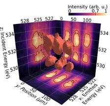
Figure 1: This visualisation of a 3D dataset demonstrates the rich information which can be obtained with a single photon energy scan (Credit: Jan Schunck (DESY), both figures: © 2021 Optical Society of America).
")
")
A collaboration coordinated by scientists from DESY has coupled imaging to X-ray absorption spectroscopy (XAS) and resonant inelastic X-ray scattering (RIXS) to study quantum materials. For the design of future functional devices, like novel computers or data storage elements, it is necessary to structure these materials on the nanoscale. Although soft X-ray spectroscopy has substantially advanced our understanding of the functionality of quantum materials, methods like XAS and RIXS are less suited for such tiny structures.
‘Some materials spontaneously form tiny domains with different functionality, for example different resistivity. Small changes in the network of these domains can make the whole material turn from an insulator to a conductor. With soft X-ray RIXS, we could not see these domains so far’, says Jan Schunck, a PhD student at DESY and the lead author of the publication in Optica. If the focus spot on the sample is larger than the structure size, the measured response is an average of potentially distinct signals.
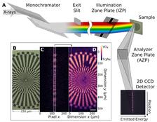
To provide a solution for this challenge, a new experimental setup was explored at the PETRA III beamline P04 (Fig. 2A) which aims at combining the RIXS technique with imaging. An off-axis Fresnel zone plate is used as an analyser to disperse emitted X-rays for the energy analysis in one dimension of a two-dimensional detector. In the orthogonal dimension the zone plate images the sample surface, thus providing separate spectra for distinct points of the sample. When the slit of the beamline monochromator is open, a line on the sample is illuminated with slightly different energies. At the same time, emission spectra are recorded from the different points on the sample. Since this method uses the full undulator beam, a spectroscopic 3D data set can be measured quickly. ‘Usually this spoils the energy resolution and burns the sample, but both problems are overcome in this setup. The method is enabled by using the great zone plates produced by our partner group around Christian David at the Paul-Scherrer Institute (PSI) in Switzerland', explains Martin Beye, group leader of the involved DESY team.
The new setup can be used for 2D fluorescence imaging as well by recording the X-ray emission from each point under illumination of the line focus (Fig. 2C) and subsequently scanning the sample in the perpendicular direction. A ‘Siemens star’ test pattern (Fig. 2B and 2D) was used to determine the spatial resolution of the setup which is 1.8 µm. Furthermore, to explore the possibilities for combined imaging and spectroscopy, partners from University Kiel produced structured VO2 thin-film samples. The structures were squares of 30 µm edge length with 10 µm spacing. With small energy scans, a 3D dataset was recorded that combines incident and emitted energy, as well as spatial information (see Fig. 1).
This novel combination of imaging and spectroscopy could be brought to a new level with PETRA IV, where a larger instrument would benefit from the dramatic increase in brightness and allow for improving energy and spatial resolution at the same time. This could provide extraordinary insights into the functionality of quantum materials on the microscopic scale.
Reference:
J. O. Schunck, F. Döring, B. Rösner, J. Buck, R. Y. Engel, P. S. Miedema, S. K. Mahatha, M. Hoesch, A. Petraru, H. Kohlstedt, C. Schüssler-Langeheine, K. Rossnagel, C. David, and M. Beye, Soft X-ray imaging spectroscopy with micrometer resolution, Optica 8 (2021), DOI: 10.1364/OPTICA.405977