


")
")
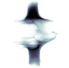
The water jet at 20 nanoseconds after being struck by the infrared laser (Credit: DESY, Johannes Hagemann et al.).

")
")
An interdisciplinary team of scientists, including from DESY, has used ultrashort X-ray pulses to image extremely rapidly exploding jets of water. The experiments, carried out at the X-ray laser facility European XFEL, aim to study ultrafast events that occur in a very small region by means of X-ray holography. “As an example of such an event, we opted for a fine water jet which is made to explode at a point along its path by an infrared laser,” says DESY’s Johannes Hagemann, lead author of the study that has been published in the Journal of Synchrotron Radiation.
“In order to record a process of this kind, the light pulse needs to be much shorter than the process itself,” Hagemann explains. “Otherwise, the image will be blurred by the relative motion of the object under scrutiny.” The European XFEL generates X-ray pulses that last only a few tens of femtoseconds. A femtosecond is a millionth of a billionth of a second. At the MID (Materials Imaging and Dynamics) beamline, the researchers shone the X-ray at a jet of water that was just 0.04 millimetres across as it was struck by a powerful infrared laser. The infrared light from the laser abruptly heats the water jet at one point, causing it to evaporate there within approximately 20 nanoseconds. A nanosecond is a billionth of a second.
The resulting images reveal the detailed dynamics of the exploding water jet. “To obtain these images, we had to overcome two obstacles,” explains Hagemann, who works in the group of Christian Schroer, Lead Scientist at DESY and one of the co-authors of the paper. “For one thing, the X-ray pulses do not provide a steady level of illumination; it is constantly fluctuating. For another, we initially only get holograms rather than real images, because the lenses available for X-rays are not of such high quality as lenses for visible light, like the ones that produce the image in a camera, for example.”
To overcome the first obstacle, the scientists took numerous images with the fluctuating lighting levels and used these to develop a mathematical model. “Using this, it is possible to retrospectively describe the illumination during any measurement,” says Hagemann. “Only then can the second obstacle be overcome.” The holograms obtained with the help of the illumination model have to be reconstructed numerically to produce the actual image of the water jet and its explosion. “Although this means doing some extra work to begin with, ultimately it pays off,” explains Hagemann. “Because the images we obtain are not simply pictures, but a measurement of the electron density of the imaged object. This allows us to identify regions of higher density, for example, which occurs in a shock wave. In addition, the density allows other physical quantities to be deduced, such as the pressure or temperature.”
The exploding water jet is not just a model system, though; it also has practical significance. On the one hand, fast evaporation by short laser pulses is being used for medical surgery; on the other hand, water jets are often used to carry biological samples, such as protein crystals, into the path of an X-ray laser beam, in order to explore their structure. This experiment has now shown that such fine water jets are also suitable for carrying larger objects, such as intact, live cells, into the X-ray beam in order to examine them. The advantage of using a water jet is that the cells remain in an aqueous environment as in the body. They do not have to be immobilised or dried.
The successful imaging with the X-ray laser opens up numerous new possibilities for investigations. “In the future, we want to use this imaging technique to capture other high-speed processes also in biological and soft matter in water,” says principal investigator Tim Salditt from Göttingen University.
This study is the first scientific publication to report experiments carried out at the recently opened MID instrument of the European XFEL, whose head Anders Madsen is also one of the authors. Researchers from the Universities of Göttingen and Hamburg, from the European XFEL and from DESY were involved in the work. DESY is the principal shareholder of the European XFEL, which begins on the DESY site in Hamburg and runs all the way to the neighbouring town of Schenefeld, in Schleswig-Holstein.
(from DESY News)
Reference:
Single-Pulse Phase-Contrast Imaging at Free-Electron Lasers in the Hard X-ray Regime; Johannes Hagemann, Malte Vassholz, Hannes Hoeppe, Markus Osterhoff, Juan M. Rosselló, Robert Mettin, Frank Seiboth, Andreas Schropp, Johannes Möller, Jörg Hallmann, Chan Kim, Markus Scholz, Ulrike Boesenberg, Robert Schaffer, Alexey Zozulya, Wei Lu, Roman Shayduk, Anders Madsen, Christian G. Schroer and Tim Salditt; Journal of Synchrotron Radiation, 2020; DOI: 10.1107/S160057752001557X