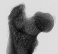
Femur transmission image.

One of the key strengths of hard x-rays is their large penetration depth in matter, allowing one to investigate the interior of an object without destructive sample preparation. Hard x-ray microscopy takes advantage of this. In combination with tomography, hard x-ray microscopy yields the three-dimensional inner structure of an object with high spatial resolution.
In addition, there is a variety of x-ray analytical techniques that can be used as contrast in x-ray microscopy. For example, using x-ray fluorescence, the distribution of chemical elements can be determined inside a sample. X-ray diffraction can be used to gain information about the local nano-structure of a specimen down to atomic resolution. Absorption spectroscopy yields the chemical state and the local chemical environment of a given atomic species.
Mainly two types of x-ray microscopy can be distinguished: full-field and scanning microscopy. While these techniques work best at synchrotron radiation sources, they can also be implemented in the home laboratory using a microfocus x-ray source.


