


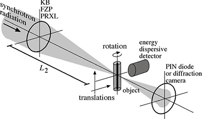
Scanning microscopy scheme.
In scanning microscopy, a small but intensive x-ray beam is used to scan the sample. This microbeam is typically generated by an x-ray optic. At each position of the scan, an x-ray analytical signal, such as the x-ray fluorescence, absorption spectroscopy, or diffraction, from the sample is recorded. In this way, elemental, chemical, and (nano-)structural information from the sample can be obtained.
")
")
Here are some examples of x-ray scanning microscopy and tomography:
- fluorescence microtomography of a root of the mahogany tree,
- tomographic absorption spectroscopy of a catalyst inside a reactor capillary,
- tomographic small-angle x-ray scattering of an injection molded polyethylene rod.