


Full-field imaging scheme.
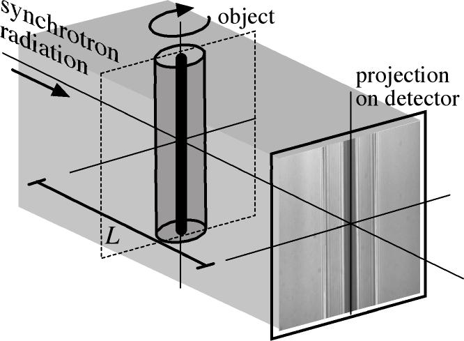
The simplest form of x-ray microscopy is done in projection. At a synchrotron radiation source, this is typically done in parallel projection as shown in the figure on the left. At the microfocus tube, magnifying projection imaging is most often used.
A more advanced form of full-field microscopy is done with the help of an x-ray optic that is used as objective lens. The sample is illuminated from the back by hard x-rays and imaged by an objective lens onto a two-dimensional detector. The illumination is usually optimized with the help of a condensor lens (not shown). As x-ray optic, Fresnel zone plates and refractive x-ray lenses can be used.

Full-field imaging in magnified geometry.