


")
")
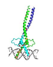
Resolved structure of the Microphthalmia-associated Transcription Faktor MITF with the characteristic kink. Mouse mutations (with pigmentation phenotypes) are colored in orange, and human mutations found in Tietz or Waardenburg syndrome are colored in red. Credit: Vivian Pogenberg/EMBL Hamburg
With the X-ray vision of DESY's light source DORIS, a research team from Hamburg and Iceland has uncovered the molecular structure of a master regulator central to the most deadly form of skin cancer, melanoma. The results, published in the scientific journal "Genes & Development", throw new light on the workings of the so-called Microphthalmia-associated Transcription Factor MITF, that is not only connected to skin cancer, but also to a variety of hereditary diseases where the production of the skin pigment melanin is disturbed, and to certain aspects of ageing. "Our data could provide a rational basis for the development of tailor-made drugs targeting MITF", explains first author Vivian Pogenberg from the Hamburg branch of the European Molecular Biology Laboratory (EMBL).
Melanoma is a malignant tumor of the cells that produce the skin pigment melanin, the melanocytes. It is not the most common form of skin cancer but the one with the greatest death toll by far: about 3 out of 4 skin cancer related deaths are caused by melanoma. Important for the development of melanoma are malfunctions of the Microphthalmia-associated Transcription Factor MITF. Transcription factors regulate which part of the DNA is read and transcribed into a blueprint for a protein within the cell. Only few parts of the DNA are active in each cell, and this activity also changes with time. MITF for instance activates the cell's machinery to turn the amino acid tyrosine into the pigment melanin.
But MITF also makes stem cells turn into melanocytes in the first place and controls cell proliferation and death in these cells. That's why MITF is called a master regulator. In fact, it also has functions in other cell types like mast cells of the immune system and bone eating osteoclasts. Mutations in MITF not only play a role in the development of skin cancer, but also cause severe genetic diseases like the Tietz and Waardenburg syndromes that lead to deafness, skin and hair pigmentation defects, abnormal eye anatomy and altered vision. The transcription factor also plays a role in our hair turning grey with age and other age-related pigmentation alterations.
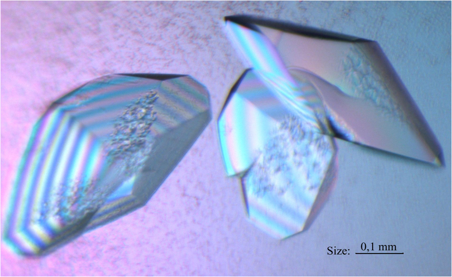
The researchers crystallised MITF in the lab and x-rayed them with DORIS. Crystals scatter X-rays in characteristic ways and produce diffraction patterns from which the structure of the crystal - and here MITF - can be reconstructed. The analysis revealed unexpected molecular insertions that give MITF a unique kink. MITF forms a dimer with a long coiled-coil protein “zipper”, and the kink in this zipper limits MITF's ability to bind to other transcription factors. The team could also identify structural changes caused by a number of MITF mutations known to lead to particular coat colours in mice and to Tietz or Waardenburg syndrome in humans. The different forms of MITF were supplied by the University of Iceland, where the lab of Eiríkur Steingrímsson hosts a comprehensive MITF library. Steingrimsson also provided his expertise in cell biology and genetics to support the structural data produced in Hamburg.
Thanks to the structural information from DORIS the team could also investigate the binding site of MITF to the DNA at the European Synchrotron Radiation Facility (ESRF) in Grenoble, France. The analysis revealed for instance that the eponymous mutation (the one leading to white coats and small eyes - or microphthalmia - in mice) causes structural changes in the MITF that prevents it from binding to the DNA. Other mutations also affect the binding site to the DNA, making MITF bind to the wrong genes. "Ultimately the goal will be to fully understand how MITF functions to evaluate how it can be targeted for potential treatment", says Matthias Wilmanns, a group leader at EMBL Hamburg. "One way would be, for instance, to design molecules that specifically stop MITF dimerisation in melanocytes," explains Pogenberg. "Or, on an alternative route, a different custom made molecule could stop the recognition of DNA by MITF."
Reference:
"Restricted leucine zipper dimerization and specificity of DNA recognition of the melanocyte master regulator MITF"; Vivian Pogenberg et al.; "Genes & Development", 2012; DOI: 10.1101/gad.198192.112
"Restricted leucine zipper dimerization and specificity of DNA recognition of the melanocyte master regulator MITF"; Vivian Pogenberg et al.; "Genes & Development", 2012; DOI: 10.1101/gad.198192.112