





The uterus is supported by several ligaments, the largest of which was analysed using PETRA III’s X-rays by the research team for the quality of its collagen content with respect to a person’s age. (Picture: “Utérus” by Servier Medical Art, CC BY 2.0)

Women’s health is underrepresented in the medical canon. Case in point: pelvic organ prolapse will affect about 40% of all women, mostly toward the latter half of their lives, and yet little is known about why it happens or how to treat it. A team of scientists from Austrian and German institutes has taken an important step towards improving that understanding – and they used PETRA III at DESY for it. Using samples of ligaments surrounding the uterus, the research team, led by scientists from the Medical University of Graz and the University Medical Center Hamburg-Eppendorf (UKE), tested a postulated reason for prolapse: they compared the behaviour of the structural protein collagen across women of various ages. The results, published in the journal Acta Biomaterialia, help to understand the role of the ligaments for uterine support.
Any sort of prolapse occurs when an organ shifts out of its healthy position in the body. For pelvic organ prolapse, medical researchers have hypothesised that the cause could be the weakening of the ligaments around the uterus, one of which is the broad ligament. “The broad ligament connects the uterus to the inner pelvic wall,” says Annika vom Scheidt, she is a scientist at the Medical University of Graz and leader of the research team. Ligaments are sturdy but stretchable connective tissues between bone and muscle or organs and their surroundings. Their strength and elasticity both come from the protein collagen, a biomolecule that exists in large triple-helix strands of amino acids. “Collagen is the load-bearing protein in human connective tissue, and it regulates how much tissue can stretch under load,” vom Scheidt says.
Since ligaments are rich in collagen, the hypothesis was that the ligaments fail because of age-related degradation of the protein. To test this, the team examined uterine ligaments from women of a wide range of ages – between 18 and 89. The team of engineers, physicists and clinicians decided to use PETRA III’s X-rays to examine the state of the collagen in the ligaments. They used experimental station P03, which examines interfaces of functional materials, from solar cells to novel materials – or biological matter. “We can also scan bones, muscles, implants and many other samples that have direct medical relevance as well as an impact,” says Matthias Schwartzkopf, scientist at the PETRA III beamline P03.
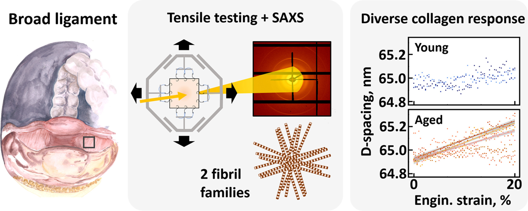
In this case, the team used submillimetre-thin segments of ligament tissue as their sample, comparing the younger and older tissues while also adding biaxial tensile stress to the tissue. Upon scanning for the structure of collagen, they found something surprising. “There was little to no difference between the collagen of younger women and older women,” Schwartzkopf says.
“Aging alone may not degrade collagen structure,” says vom Scheidt. “Our results show no clear age-related changes in the collagen behaviour of the broad ligament but a subset of older women show subtle, nanoscale changes in collagen that could contribute to disease development.” These findings provide mechanistic insight that is still largely missing and may contribute to better future treatment strategies by informing surgeons about tissue quality.
Schwartzkopf emphasises the importance of body donations in the success of this study. “This was only possible because people requested to have their bodies donated for teaching and research – for this and a lot of medical research, these contributions are vital and help make improvements in many other people’s lives possible.”
“Understanding whether the biomechanical behaviour of the support tissues of the uterus change with age is essential to improve treatment possibilities for pelvic floor disorders,” says vom Scheidt.
(Partly rom DESY News)
Original publication:
A. vom Scheidt et al., "Collagen behavior in the human broad ligament under biaxial tensile testing with small-angle X-ray scattering," Acta Biomaterialia (2026), DOI: 10.1016/j.actbio.2026.04.050
