


")
")
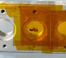
One of the sample holders used in the experiment holding a blood clot. (Photo: DESY)
")
")
Heart lung machines are used to save life but also come with risks. An international research team led by the Swedish Royal Institute of Technology (KTH), using X-ray measurements at PETRA III alongside simulations and microscopy, has uncovered how blood flow directly shapes the structure of blood clots. The study was carried out by a collaboration between KTH, Karolinska Institute, Umeå University and DESY and has been published in Scientific Reports.
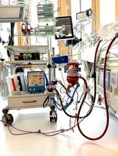
Extracorporeal membrane oxygenation (ECMO) is used when a patient’s heart or lungs temporarily fail. In this therapy, blood is circulated through an external circuit where it is oxygenated before being returned to the body. ECMO is a last resort in critical care and can be life-saving in cases of severe respiratory or cardiac failure.
However, conditions inside these systems differ markedly from those in the body. Blood is exposed to high-mechanical forces and regions of trapping flow. These conditions can increase the risk of complications such as clot formation, bleeding or damage to red blood cells. Understanding how these clots form is therefore essential for improving both devices and treatment strategies.
To address this, the research team analysed two blood clots taken from separate ECMO circuits used in two patients. The aim was to understand how flow conditions inside the circuits influence the structure and development of clots.
The team combined several complementary methods. Computer simulations of blood flow identified regions in the system where clot formation is more likely. These simulations highlight areas with particularly high mechanical stress or complex flow patterns which are considered critical for the initiation of clots.
To examine the clots themselves, the researchers used X-ray scattering at the PETRA III beamline P03 at DESY. This technique allows scientists to probe the internal structure of a sample. Scattering measurements revealed the density and alignment of fibrin fibres which form the structural backbone of blood clots. Electron microscopy provided additional surface detail on how blood cells and fibres are arranged at the microscopic scale.
Only by combining these approaches was it possible to establish a direct link: The local flow conditions within the system are reflected in the internal structure of the blood clots. Mechanical forces therefore influence not only whether a clot forms but also how it is structured and grows — findings that are also highly relevant for the development of safer medical devices.
“Understanding how mechanical forces drive clot formation and growth in these extreme environments is essential for improving ECMO system design and ultimately enhancing patient safety,” says Frida Nilsson, corresponding author of the study and researcher at KTH.
“The X-ray measurements at PETRA III allow us to look inside these clots and quantify how their internal structure is organised at very small scales,” adds DESY scientist Stephan V. Roth. Nilsson continues: “This information is very difficult to obtain otherwise and is essential for connecting flow conditions to the material properties of the clot. This work forms part of a broader research effort led by Professor Lisa Prahl Wittberg at KTH and Associate Professor Lars Mikael Broman at Karolinska Institute, aimed at improving ECMO device design and optimising operation to reduce complications and improve patient outcomes.”
The findings could help to make life-support systems safer and reduce complications for patients. With next-generation sources such as PETRA IV, this approach could be pushed further, allowing researchers to study clot structure at even finer scales and gain a more detailed picture of how flow shapes their internal organisation.
(Partly from DESY News)
Reference:
Nilsson, F. et al., Multimodal characterization of flow-induced thrombus initiation and growth in extracorporeal membrane oxygenation. Scientific Reports (2026), DOI: 10.1038/s41598-026-40177-3