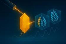
Conceptual illustration based on a user-defined concept for time-resolved cryo-EM and X-ray crystallography (generated with the assistance of ChatGPT (OpenAI, 2025)).
An interdisciplinary research team from the Center for Free-Electron Laser Science (CFEL) at DESY, the Centre for Structural Systems Biology (CSSB), the Center for Ultrafast Imaging (CUI) at the University of Hamburg and Uppsala University has published a review in the renowned journal Nature Methods that sheds light on the future potential of combining two key technologies in structural biology: X-ray imaging and cryo-electron microscopy (cryo-EM). By combining ideas and approaches developed separately in these two fields, the researchers highlight new ways to visualise biological processes in real time and high resolution – a promising approach for the development of future molecular films and personalised drugs. How do molecules move in living cells? How do they interact with each other? How can these processes be visualised and controlled? These are central questions in structural biology and physics. Although various methods already allow insights into molecular dynamics, it is difficult for researchers to visualise structural changes with high temporal and spatial resolution. “Biological functions are based on movements of molecules, atoms and electrons that take place over different time scales,” explains Amir Banari, a researcher at CUI and first author of the current study. “Our team is developing new methodological approaches for this – in particular by combining cryo-electron microscopy and X-ray imaging.”
Making moving molecules visible
Time-resolved structural biology aims to capture biological molecules not just as static structures but in motion – thus gaining important insights into how they function. The biggest challenge with this research approach is that many of these reactions take place extremely quickly and conventional imaging methods often only capture stable states. At the same time, the intense radiation used for the investigation causes damage to the sensitive samples. To overcome these hurdles, the team relies on the targeted combination of two complementary methods of structural biology: cryo-electron microscopy and X-ray crystallography. In their publication they were able to show how the two complement each other perfectly. “With our work we are building a bridge between these previously separate methods,” says DESY researcher Dominik Oberthür. ” New preparation methods, serial data collection and AI-supported analyses are key to combining the strengths of both worlds.”
Interdisciplinarity as a success factor
A central element of the project is the cooperation between physics, biochemistry and data analysis. With the support of CUI, experts from various disciplines were brought together to network the existing technologies on the research campus. Multimodal imaging and AI-supported analysis methods make it possible to investigate biological processes in space and time with unprecedented resolution. “Only through interdisciplinary collaboration can we create new tools to better understand the dynamics of biological processes and make them usable for medical purposes in the long term,” emphasises Carolin Seuring from the CSSB.
Looking to the future: tailor-made drugs, PETRA IV, and AI-supported analyses
Findings from time-resolved structural biology and synergies between cryo-EM and X-ray sciences promise a wide range of applications, such as the development of drugs with a more targeted effect and fewer side effects, advances in personalised medicine through tailored therapies, as well as innovations in biotechnology and energy research. “By understanding how biomolecules change structurally during reactions, we can, for example, develop drugs that specifically target transient or intermediate states,” explains Anna Munke, co-author of the study.
The research group plans to further improve the integration of the techniques of cryo-EM with X-ray imaging and diffraction at X-ray free-electron laser and synchrotron facilities. The use of AI in data analysis should enable the consolidation of information from disparate measurements to improve the structural determination of volatile intermediate states. With the next-generation synchrotron – PETRA IV at DESY – the team expects a technological leap forward. Closer collaboration between structural biology and cryo-electron microscopy will open up further possibilities: “Combining measurements from PETRA IV, the European XFEL and cryo-EM will allow us to capture larger ranges and finer details of dynamic structural changes with higher contrast,” says DESY lead scientist, CUI researcher and University of Hamburg professor Henry Chapman. “Our long-term goal is to visualise even complex processes in living cells with unprecedented resolution.”
(from DESY News)
Reference:
Advancing time-resolved structural biology: latest strategies in cryo-EM and X-ray crystallography; A. Banari et al., Nature Methods (2025), DOI: 10.1038/s41592-025-02659-6


