


Study of clusters
Low energy spectroscopy
Radiation in the VUV region of the spectrum (10-100 eV) is suited to several applications: Elucidation of atomic bonds, study of correlations between electrons and of the dynamics of chemical reaction pathways. UV or visible light can be used for the quantitative description of solutions of transition metals or highly conjugated organic compounds
The lowest energies in this category are in the IR or Terahertz region of the spectrum (< 1eV). In IR spectroscopy the activation of vibrational and other modes of excitement of atomic bonds generates dips (also called "peaks") in an absorption spectrum, which can be used to analyze bonding in organic compounds.
Terahertz radiation is useful for the study of biological systems since it is non-ionizing and thus doesn't damage tissue, but can still penetrate many materials.
Generally the techniques work by illuminating a sample (solid, gaseous) and deducing information about it by analyzing the product particles, which can be photons, electrons, ions, atomic clusters, etc. An example would be the analysis of the transmitted photons in the form of an absorption spectrum. Typical applications are in:
- Complex materials
- Surfaces, Clusters
- Atomic& molecular physics, astrophysics
- Combustion, chemical dynamics
- Biological systems
The high flux and brightness achieved in modern light sources allow the use of very advanced analysis tools. High signal rate and resolving power can be achieved.
Includes:VUV, IR, photo-ion, THz, FTIR, PEPICO spectroscopy; PES; ARPES; UPS; COLTRIMS
Soft X-ray spectroscopy
By exciting electrons in shallow core electrons (100-2000 eV) the electronic structure of material can be analyzed.
In all techniques in this category a specific property of a material is analyzed while sweeping the photon energy through a range of values. A simple way of analysis is just measuring basic properties of the sample like absorption, reflection, transmission and reflection at the different photon energies. Absorption spectroscopy is the most widely used example.
“Double spectroscopy” allows more sophisticated investigation.
“Photon-in/electron-out” spectroscopy encompasses two techniques:
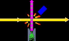
In photoelectron spectroscopy (XPS) the incident x-ray photon removes a core-level electron from an atom, whose energy is then measured. Together with the energy of the incident photon this then yields information about how tightly the electron was bound and thus about e.g. oxidation state and bonding.
In Auger spectroscopy an electron from a higher energy level fills the generated vacancy and in the process transfers its excess energy to a neighboring electron which is then emitted and analyzed.
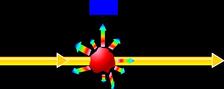
“Photon-in/photon-out” spectroscopy or x-ray fluorescence (XRF) is similar to Auger spectroscopy. The difference is that when the electron fills the vacancy it releases its energy in the form of a photon, which is then studied.
Some chiral and magnetic systems also respond to circular polarization of the light, meaning this is another tool of investigation. The method employed is x-ray circular dichromism (XMCD). By comparing images taken with right and left circular polarized light, properties like spin and orbital magnetic momentum can be analyzed.
Applications include:
- Complex materials
- Magnetic materials
- Wet samples at ambient pressure (environmental science)
- Catalysis
The great brilliance of modern light sources makes the photon-in/photon-out techniques possible, where the low collision cross-section usually does not give enough collisions. To adapt the technique to different elements with different energy levels, the tunability of the synchrotron radiation is essential.
Includes: XAS; NEXFAS, Auger spectroscopy; SXES; RIXS; XMCD; XPS;XRF
Hard X-ray spectroscopy
Since this technique is element-, oxidation-state- and symmetry-specific due to the differing core-level energies, it finds application in a wide range of fields to analyze geometric and electronic structure. It is useful in the characterization of new materials and elucidation of very dilute chemicals, e.g. for environmental reasons.
By measuring the intensity of incident and transmitted radiation while sweeping through element-specific photon energy levels corresponding to the core electrons’ energies (“edges”), analysis of the sample is possible. Techniques include:
EXAFS (extended x-ray absorption fine structure): A wide sweep of radiation energy above an “edge” displays small oscillations in the absorption. From this, information about nearest neighbor atoms can be deduced.
NEXAFS (Near-edge x-ray absorption fine structure): A more narrow sweep generates a “fingerprint” of characteristic peaks in the absorption spectrum for the specific arrangement of atomic bonding around the atom of origin. (Same as XANES)
Mössbauer Spectroscopy: Using gamma rays of narrowly defined energy and mapping their absorption as a function of energy yields information about the chemical environments of targeted nuclei and allows characterization of the sample.
The technique is based on the same mechanisms as described in soft x-ray spectroscopy. Only now not the originally ejected photoelectrons or Auger electrons are studied, but the ones that were inelastically scattered afterwards. In each case the oscillations in the spectrum are generated by interference between the wave associated with the originally ejected electron and electronic wavelets backscattered from other atoms.
Again the tunability of the radiation is essential for the techniques, and its intensity makes the study of dilute samples possible.
Includes: EXAFS, NEXAFS, XANES, Mössbauer spectroscopy; XAS; XMCD