


")
")
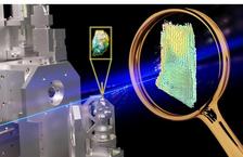
Experimental setup with tissue on the sample holder (middle), 3D vector field of the collagen direction and degree of orientation obtained by SAXS-Tensor-Tomography (right). (Figure: S. Haas, DESY)
A collaborative effort between researchers from DESY, the University Medical Center Hamburg-Eppendorf (UKE), Chalmers University in Sweden and the Paul Scherrer Institute in Switzerland has yielded a cutting-edge multimodal imaging approach to investigate breast cancer tissue. With the help of this technique, researchers can simultaneously extract information about the nanostructure of the tumor and quantify the chemical elements present in a millimeter-scale sample in all three dimensions. A unique combination of research possibilities at PETRA III and new analysis methods enables this high level of detail.
Breast cancer caused 685 000 deaths globally in 2020 according to the WHO. It is not life-threatening in its earliest form. But if the cancer cells are able to spread further in the tissue to nearby lymph nodes or important organs, this metastasis can be fatal. In a recent pilot study published in Nature Scientific Reports, the team applied this revolutionary imaging approach to a breast cancer sample. The results show how key molecules collectively influence the metastatic mechanism. This breakthrough paves the way for an in-depth investigation of breast cancer metastasis, promising novel therapeutic approaches and personalised treatment strategies, which could ultimately improve patients’ lives if recognized early enough.
Traditional experimental models often fall short, relying on 2D cell cultures or animal models that do not faithfully replicate the complex physiological patterns of human tumor environments. The multimodal imaging approach presented in this study represents a significant step forward by providing simultaneous nanoscale morphological and physiological information from real samples, thus giving researchers information about the shape and composition of real cancer tissue.
André Conceição, the first author and beamline scientist at the PETRA III SAXSMAT beamline P62, emphasises, "Although demonstrated for breast cancer, this approach's versatility extends to other organs and diseases."
The study opens avenues for further exploration of breast cancer metastasis and pre-metastatic niches (PMNs). Advanced X-ray multimodal tomography can generate complementary 3D maps for different breast cancer molecular subtypes. It holds the potential to contribute to the development of more targeted and effective strategies for diagnosis and treatment.
Volkmar Müller, professor of gynaecology at the UKE and co-author of the study, underlines the importance of understanding the 3D architecture of the tumor microenvironment. Where the tumor is located in the patient’s tissue and how it is embedded, is a key for the development of further novel therapeutic approaches to interrupt the metastatic mechanism. For this one of the most striking features is collagen in the direct vicinity as well as the concentration of iron and zinc. Malte Mohme, lecturer on CNS tumor immunology at the UKE and co-author of the study, explains: "The work by Conceicao et al. highlights the critical role of metastasis in neuro-oncology, focusing on the systemic spread of breast cancer. By using advanced multimodal X-ray computed tomography, the study reveals how changes in the extracellular matrix, particularly through zinc and iron accumulation, might influence metastasis. Insights into matrix metalloproteinases (MMPs) and collagen orientation offer a new understanding of cancer cell migration and brain tumor-homing, a complex, multistep process by cells to travel from a distance to a tumor. This knowledge is vital for neuro-oncology, as it opens pathways for targeted therapies to disrupt metastatic processes. The findings underscore the importance of the detailed composition of the extracellular matrix in understanding metastasis, potentially leading to improved outcomes in patients with brain metastases."
Sylvio Haas, beamline manager of the PETRA III beamline P62 and co-author of the study, highlights the unique experimental setup using small-angle X-ray scattering tensor tomography (SAXS-TT) and X-ray fluorescence computed tomography (XRF-CT). Higher-resolution images will be possible in the future, in particular with PETRA IV, the upgrade of PETRA III. While the proposed imaging approach currently relies on state-of-the-art instrumentation and substantial resources, André Conceição is optimistic about further developments of the setup. Anticipated advancements in instrumentation and machine learning hold the potential to expedite data acquisition and enhance the 3D reconstruction process, making the innovative imaging technique applicable to a broader range of laboratories and clinical settings. This research not only advances the understanding of breast cancer metastasis but also emphasis the relevance of developing novel therapeutic strategies in neurosurgical oncology.
(Partly from DESY News)
Reference:
A. L. C. Conceição, V. Müller, E.-C. Burandt, M. Mohme, L. C. Nielsen, M. Liebi, S. Haas, Unveiling breast cancer metastasis through an advanced X-ray imaging approach, Sci Rep (2024) DOI: 10.1038/s41598-024-51945-4