


")
")
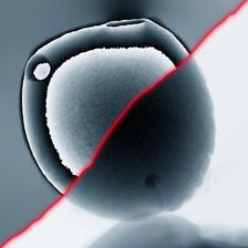
Detail from figures below: Far-field ptychography of a micro meteorite recorded at PETRA III. Left upper part: phases of the conventional ptychography reconstruction. Right lower part: phases of the refractive reconstruction. (Figure from original publication).
")
")
")
")
In X-ray microscopy, it is often the optics that limit spatial resolution or cause distortion in the image. A radical way to get around this problem is to omit the optics altogether. The price of this ‘lensless’ microscopy is that the image must be calculated from the intensity pattern formed on the detector by the light scattered from the sample. But the calculation yields more than just an image: It provides a quantitative measure of the refraction and attenuation inside the studied object. In this way, even very transparent objects obtain a strong contrast, for example making the method well suited for visualising unstained biological cells and tissues.
So far, recovering the image from the scattered intensity included some ambiguity: the refraction inside the object advances the X-ray wave front. Once this shift of the wave front reaches the wavelength, the numerical image reconstruction can no longer distinguish this wave front from an unshifted wave. Therefore, the effect of refraction could only be unambiguously obtained with the conventional methods if it was small enough. In all other cases, this phase shift had to be corrected by shifting the wave by the correct integer multiple of the wavelength. This so-called ‘phase unwrapping’ can be difficult to achieve but is needed to get quantitative information about the sample.
A team, headed by Christian Schroer from DESY, has developed a new approach to directly reconstruct the refraction inside the sample and avoid the phase wrapping problem altogether. With this new refractive framework, the physical properties of a sample can be quantitatively reconstructed avoiding all difficulties previously related to phase unwrapping. The mathematical approach is quite general, so it can be applied to a large class of problems commonly encountered in science and engineering. The team applied them to two different X-ray microscopy techniques commonly used at modern synchrotron radiation sources, such as PETRA III.
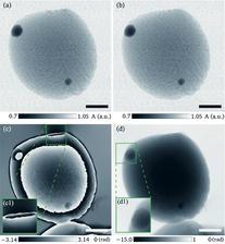
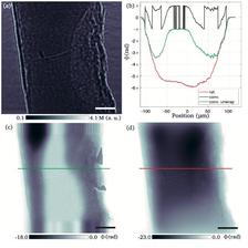
In particular, following the general mathematical scheme for the refractive framework, the team derived refractive algorithms for ptychography and holography based on existing iterative projection algorithms. They validated the two algorithms on experimental data obtained with the X-ray microscopes at beamlines P05 and P06 at PETRA III. For conventional ptychography and holography, the strongly phase-shifting samples are challenging to reconstruct, as the respective examples illustrate. Conventionally, phase wraps, especially if there are multiple, are often not accurately reconstructed and can lead to inconsistencies, such as phase vortices shown near the upper edge of the micro meteorite shown in the second figure (panel c). With the new refractive framework, this problem is negligible (second figure, panel d). In addition, the conventional holographic phase reconstruction can fail altogether when the phase shift inside the object becomes too large, as shown in the last figure. Here, the new approach is far superior, leading to a quantitative reconstruction of the object.
The refractive framework greatly simplifies the task of phase retrieval and is much more general than presented in this study. It can be applied to other lensless imaging modalities using X-rays or electrons as a probe. At future synchrotron radiation sources, such as PETRA IV, the refractive framework will open new possibilities to study even larger sample volumes at high resolution.
Original publication:
Felix Wittwer, Johannes Hagemann, Dennis Brückner, Silja Flenner, and Christian G. Schroer, Phase retrieval framework for direct reconstruction of the projected refractive index applied to ptychography and holography, Optica (2022) DOI: 10.1364/OPTICA.447021