


")
")
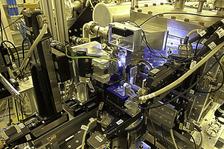
Overview of the sample area of the GINI-X setup
")
")
")
")
The Göttingen Instrument for Nano-Imaging with X-Rays (GINIX) is a dedicated endstation for coherent nano-diffraction and imaging at the Coherence Beamline P10. It is located in the second experimental hutch (EH2).
The instrument has has been designed and installed by the group of Prof. Dr. Tim Salditt ( http://www.roentgen.physik.uni-goettingen.de ) in collaboration with Dr. Michael Sprung at P10 and the PETRA III project team. A powerful Kirkpatrick-Baez (KB) mirror system and a modular system of additional optical elements can be used for beam definition, filtering and focusing with spot sizes in the range from 500 nm down to 5 nm.
Three classes of experiments are routinely carried out: scanning X-ray transmission microscopy (STXM) and nano-diffraction, ptychographic imaging and in particular cone beam propagation imaging/tomography. The later is also known as near-field or holographic imaging. A unique feature of the instrument is the combination of KB focusing with waveguide optics, resulting in unparalleled clean and coherent wavefronts ideally suited for quantitative near-field phase retrieval. More specialized modalities comprise nano-focus wide angle diffraction experiments, scanning fluorescence microscopy, coherent diffraction in reflection geometry, as well as combinations of nano-focus experiments with ultra-fast time resolution (synchronization to a bunch clock).
The construction and operation is funded by Georg-August Universität Göttingen and BMBF Verbundforschung (see below).
Instrumentation and Characteristics:
GINIX is installed on a 5-axis table (IDT), that is movable on air pads and can be exchanged with the standard CDI/XPCS set-up. At the heart of GINIX, two total reflection mirrors in Kirkpatrick-Baez geometry placed in a vacuum vessel provide a two-dimensional hard X-ray focus down to about 200 nm × 200 nm in air, with a free working distance of about 16 cm between the vessel exit window and the focal plane. The accessible energy ranges are 6.0-13.8 keV. The X-ray beam can be shaped, cleaned and filtered at different positions with slits, pinholes and waveguides. Beamstops to protect detectors can be inserted at various positions.
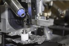
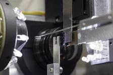
The sample is handled by a three-axis piezo on top of two rotations and large-scale stepper motors, allowing for movements of a few nanometres up to hundreds of millimetres. An air-bearing rotation for reduced wobble is installed for tomography applications. Two optical in-line microscopes can be used for visual alignment and observation of the sample. Radiation damage can be reduced using the additional option of a cryojet.
Detectors can be placed in a front (at a distance of 400-800 mm from the focus) or rear position behind the large flight path (at a distance of ~5.0 meters from the focus). The instrument is fully operated using SPEC, with essential P10 components included via Tango. All X-ray detectors (and user defined ROIs) can be used as counters by virtue of the Göttingen Live Viewer software. Fast STXM is available with on-line analysis.
Publications related to GINIX commissioning and operation (Status August 2013):
F. Döring, A.L. Robisch, C. Eberl, M. Osterhoff, A. Ruhlandt, T. Liese, F. Schlenkrich, S. Hoffmann, M. Bartels, T. Salditt, H.U. Krebs
Sub-5 nm hard x-ray point focusing by a combined Kirkpatrick-Baez mirror and multilayer zone plate
Optics Express, Vol. 21, Issue 16, pp. 19311-19323 (2013)
M. Krenkel, M. Bartels, and T. Salditt
Transport of intensity phase reconstruction to solve the twin image problem in holographic x-ray imaging
Optics Express, Vol. 21, Issue 2, pp. 2220-2235 (2013)
K. Giewekemeyer, R.N. Wilke, M. Osterhoff, M. Bartels, S. Kalbfleisch, T. Salditt
Versatility of a hard X-ray Kirkpatrick–Baez focus characterized by ptychography
J. Synchrotron Rad. 20, 490–497 (2013)
M. Bartels, M. Priebe, R.N. Wilke, S.P. Krüger, K. Giewekemeyer, S. Kalbfleisch, C. Olendrowitz, M. Sprung and T. Salditt
Low-dose three-dimensional hard x-ray imaging of bacterial cells
Optical Nanoscopy 2012, 1:10, doi:10.1186/2192-2853-1-10
C. Olendrowitz, M. Bartels, M. Krenkel, A. Beerlink, R. Mokso, M. Sprung and T. Salditt
Phase-contrast x-ray imaging and tomography of the nematode Caenorhabditis elegans
Phys. Med. Biol. 57 (2012) 5309–5323
S. P. Krueger, H. Neubauer, M. Bartels, S. Kalbfleisch, K. Giewekemeyer, P. J. Wilbrandt, M. Sprung and T. Salditt
sub-10 nm beam confinement by X-ray waveguides: design, fabrication and characterization of optical properties
J. Synchrotron Rad. (2012). 19, 227-236
A. Ruhlandt, T. Liese, V. Radisch, S. P. Kruger, M. Osterhoff, K. Giewekemeyer, H. U. Krebs, and T. Salditt
A combined Kirkpatrick-Baez mirror and multilayer lens for sub-10 nm x-ray focusing
AIP ADVANCES 2, 012175 (2012)
T. Salditt, S. Kalbfleisch, M. Osterhoff, S. P. Krüger, M. Bartels, K. Giewekemeyer, H. Neubauer, and M. Sprung
Partially coherent nano-focused x-ray radiation characterized by Talbot interferometry
Optics Express, Vol. 19, Issue 10, pp. 9656-9675 (2011)
S. Kalbfleisch, H. Neubauer, S. P. Krüger, M. Bartels, M. Osterhoff, D. D. Mai, K. Giewekemeyer, B. Hartmann, M. Sprung and T. Salditt
The Göttingen Holography Endstation of Beamline P10 at PETRA III/DESY
AIP Conf. Proc., Vol. 1365, pp. 96-99 (2011)
S. Kalbfleisch, M. Osterhoff, K. Giewekemeyer, H. Neubauer, S. P. Krüger, B. Hartmann, M. Bartels, M. Sprung, O. Leupold, F. Siewert, and T. Salditt
The holography endstation of beamline P10 at PETRA III
AIP. Conf. Proc., 1234, 433-436 (2010)

The GINIX construction and operation is funded BMBF (projects 05KS7MGA, 05K10MGA, 05K13MG4 and 05K16MG2) as well as by the Georg-August Universität Göttingen.