Retrieved complex valued illumination function.
In order to achieve high spatial resolution and to investigate extended samples with x-ray microscopy a scanning x-ray microscopy method with coherent illumination – ptychography - can be used.
The word ptychography was derived from the Greek words ptyché (πτυχή = fold) and gráphein (γράφειν = to write).
In this technique, the sample is scanned with a focused coherent x-ray beam. However, at each position of the scan, a two-dimensional diffraction pattern is recorded in the far field regime. Using a numerical reconstruction algorithm, both the object's complex transmissions function and the complex illuminating wavefield can be reconstructed from the series of diffraction patterns.

Currently, the object can be reconstructed with a spatial resolution down to approximately 10 nm. Compared to conventional scanning microscopy, where the spatial resolution is limited by the size of the beam, ptychography can resolve structures in the sample that are much smaller than the beam size. Therefore, ptychography is a promising method in comparison to other x-ray microscopy methods.
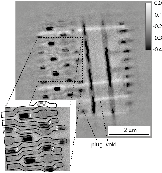
Ptychographic reconstruction of the inner life of a microchip with conducting paths.