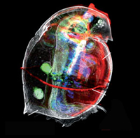
X-ray techniques reveal the metal distribution in the water flea Daphnia magna (Courtesy: JAAS, Reproduced by permission of The Royal Society of Chemistry).
Scientists from the Ghent University (Belgium), University of Debrecen (Hungary), and HASYLAB/DESY contributed to the study of tissue-specific trace level metal distributions in Daphnia magna. Daphnia magna are very small, mostly planktonic, crustaceans, and are often called water fleas. They are frequently used as organism in laboratory ecotoxicity testing and allow the quantitative investigation of the accumulation of metals within specific organs with microscopic resolution.
Studies of trace metals within organisms are a rapidly expanding research area as metals play many vital roles in biological systems. Most of the techniques currently used to analyse metal content in biological samples are destructive and give no clues about metal distribution within the system.
By combining synchrotron radiation x-ray fluorescence and laboratory x-ray absorption microtomography, L. Vincze and his colleagues were able to unravel the tissue-specific 2D and 3D metal distribution within the waterfleas without invasive sample preparation techniques. The scanning micro SR-XRF experiments were performed at Beamline L of DORIS III at DESY.
The results appeared in JAAS J. Anal. At. Spectrom., 2008, 23, 829.
(from: ...Paper and RSC News)
| Further Links |
|


