


")
")
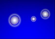
The three atoms of the excited NeKr2 trimer keep roaming around each other for up to one picosecond. (Image: FHI)
An international research team has used PETRA III to show how gas atoms rearrange themselves before they decay. In a decay process triggered by X-rays, they release low-energy electrons. The team observed how the atoms dance around each other for up to a picosecond before the system explodes. This is the first time that researchers have gained detailed insights into the temporal sequence of this decay process which also reveals associated radiation damage mechanisms.
High-energy radiation, for example in the X-ray range, can cause damage to our cells. This is because energetic radiation can excite atoms and molecules which then often decay – meaning that biomolecules are destroyed and larger biological units can lose their function. There is a wide variety of such decay processes and studying them is of great interest in order to better understand and avert radiation damage.
In a recent study, researchers from the FHI, together with international partners, investigated a radiation-induced decay process that plays a key role in radiation chemistry and biological damage processes: electron-transfer-mediated decay. In this process, one atom is excited by irradiation. Afterwards, this atom relaxes by ‘stealing’ an electron from a neighbor while the released energy ionises yet another nearby atom. The research team was able to directly track how atoms in a model system move and rearrange themselves before undergoing this exotic electronic decay process. Their work provides the most detailed real-space and real-time picture of ETMD to date.
Sophisticated combination of experiment and theory
The research team studied a simple prototype system consisting of one neon atom loosely bound to two krypton atoms (NeKr2 trimer). After ionising the neon core with soft X-rays, the researchers followed the system for up to a picosecond – an eternity on the atomic timescale – before it finally decayed by transferring an electron between neighbouring atoms and emitting a low-energy electron. Using a state-of-the-art COLTRIMS reaction microscope at the synchrotron light sources BESSY II in Berlin and the PETRA III beamline P04, the team reconstructed the molecular geometry at the exact instant the decay occurred. To interpret the measurements, fully dimensional ab initio simulations were carried out, tracking thousands of nuclear trajectories and evaluating the decay probability along each one.
Taking a movie of the non-local electronic decay
What the team discovered was striking: The atoms do not remain frozen in their initial configuration. Instead, they undergo pronounced roaming-like motion, continuously reshaping the molecular geometry and strongly influencing when and how the decay occurs.
“We can literally watch how the atoms move before the decay happens,” says Florian Trinter, one of the lead authors from FHI. “The decay is not just an electronic process – it is steered by nuclear motion in a very direct and intuitive way.”
The results reveal that ETMD does not happen from a single “preferred” structure. Instead, different molecular geometries dominate at different times: At early times, the decay occurs near the ground-state geometry, while at intermediate times one krypton atom approaches the neon atom closely and the second drifts farther away – an optimal setup for electron donation and long-range energy transfer. At later times, the system explores almost linear and highly distorted configurations, reflecting a pendular, roaming-like motion of the atoms. This dynamic reshaping leads to strongly time-dependent decay rates, varying by nearly an order of magnitude depending on geometry.
“The atoms explore large regions of configuration space before the decay finally takes place,” explains Till Jahnke from European XFEL, senior author of the study. “This shows that nuclear motion is not a minor correction – it fundamentally controls the efficiency of non-local electronic decay.”
Why It Matters
ETMD has attracted increasing attention because it efficiently produces low-energy electrons which are known to cause chemical damage in liquids and biological matter. Understanding how ETMD depends on molecular structure and motion is therefore crucial for modeling radiation damage in water and biomolecular environments, as well as for interpreting ultrafast X-ray experiments. Moreover, the current findings are very helpful for developing multiscale theoretical approaches that embed accurate decay rates into large complex systems.
By providing a detailed benchmark for the smallest system that supports ETMD involving three atoms, the present study lays the groundwork for extending these ideas to liquids, solvated ions and biological environments.
“This work shows how non-local electronic decay can be used as a powerful probe of molecular motion,” the authors conclude. “It opens the door to imaging ultrafast dynamics in weakly bound matter with unprecedented detail.”
(from DESY News)
Reference:
Florian Trinter, Jaroslav Hofierka, Jonas Rist, Max Kircher, Miriam Weller, Niklas Melzer, Dimitrios Tsitsonis, Angelina Geyer, Jan Kruse, Gregor Kastirke, Joshua B. Williams, Tsveta Miteva, Reinhard Dörner, Markus S. Schöffler, Maksim Kunitski, Nicolas Sisourat, Lorenz S. Cederbaum, and Till Jahnke, Tracking the Complex Dynamics of Electron-Transfer-Mediated Decay in Real Space and Time, Journal of the American Chemical Society (JACS), (2025), DOI: 10.1021/jacs.5c15510
Press release by the Fritz Haber Institute