


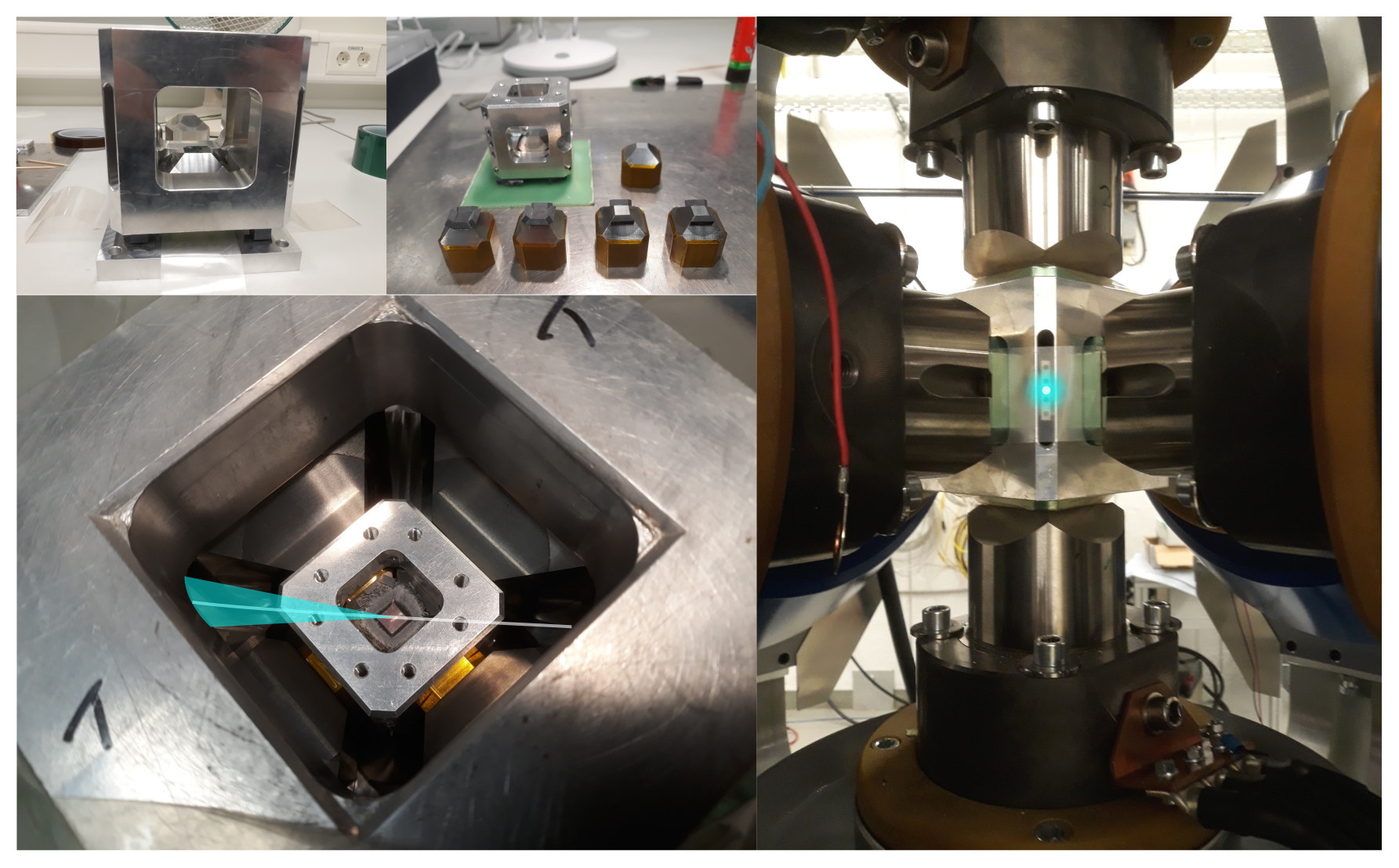
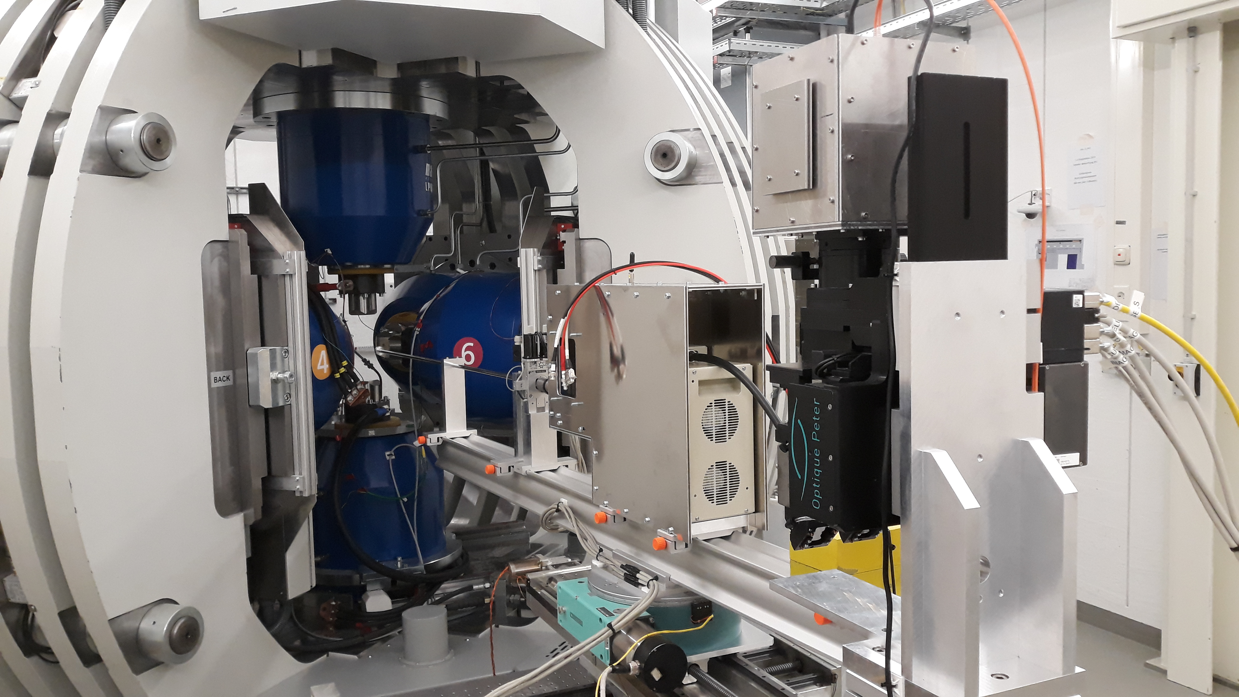
03-12-2020. The world's first discrete third stage compression geometry in a 6-ram LVP for in situ rock deformation using angle-dispersive XRD (X-rays illustrated on the photographs).
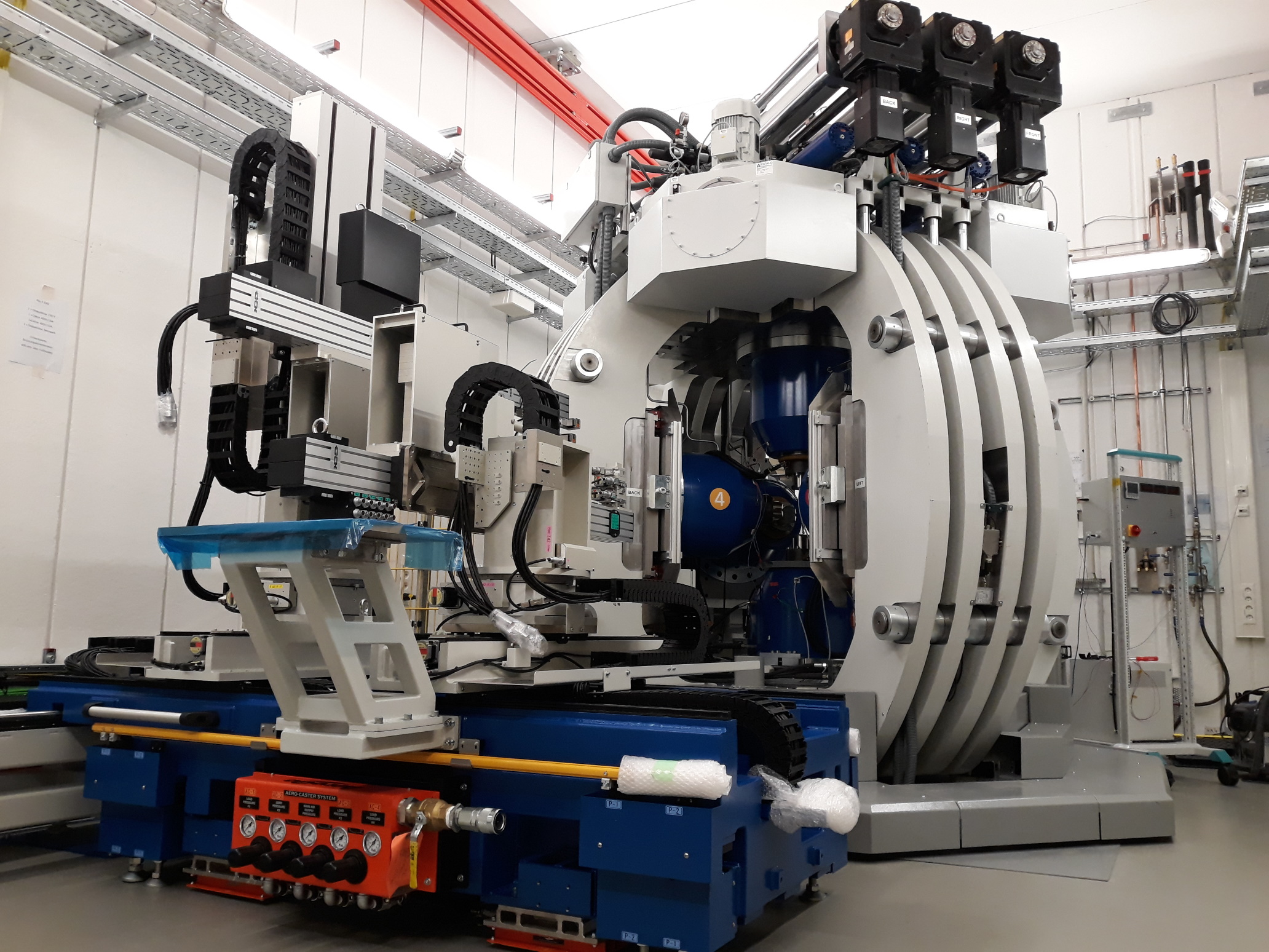
06-11-2020. The installation of the 2-detector positioning system (DPS) has started!
03-09-2020.
Tungsten wire lit by synchrotron X-rays
09-03-2020. The gauge volume for ED-XRD
Below is a calculation of the maximum length (diagonal) of the gauge volume based on the incident slit gap, the collimator slit gap and the diffraction angle. If possible, one should ensure the sample diameter is always equal or more to this value for a given geometry to avoid contribution of reflections from the sample environment.
| 2theta | Incident horz | Collimator horz | Diagonal gauge volume |
|---|---|---|---|
| (deg) | (um) | (um) | (um) |
| 4 | 50 | 30 | 1146 |
| 4 | 50 | 50 | 1433 |
| 4 | 100 | 50 | 2150 |
| 5 | 50 | 30 | 917 |
| 5 | 50 | 50 | 1146 |
| 5 | 100 | 50 | 1720 |
| 6 | 50 | 30 | 764 |
| 6 | 50 | 50 | 955 |
| 6 | 100 | 50 | 1433 |
| 7 | 50 | 30 | 655 |
| 7 | 50 | 50 | 819 |
| 7 | 100 | 50 | 1229 |
| 8 | 50 | 30 | 574 |
| 8 | 50 | 50 | 717 |
| 8 | 100 | 50 | 1075 |
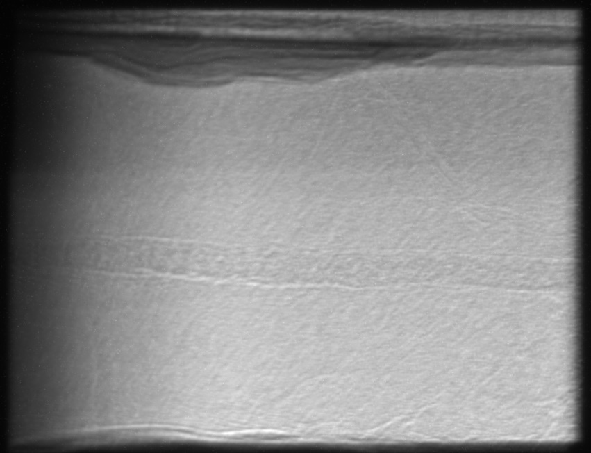
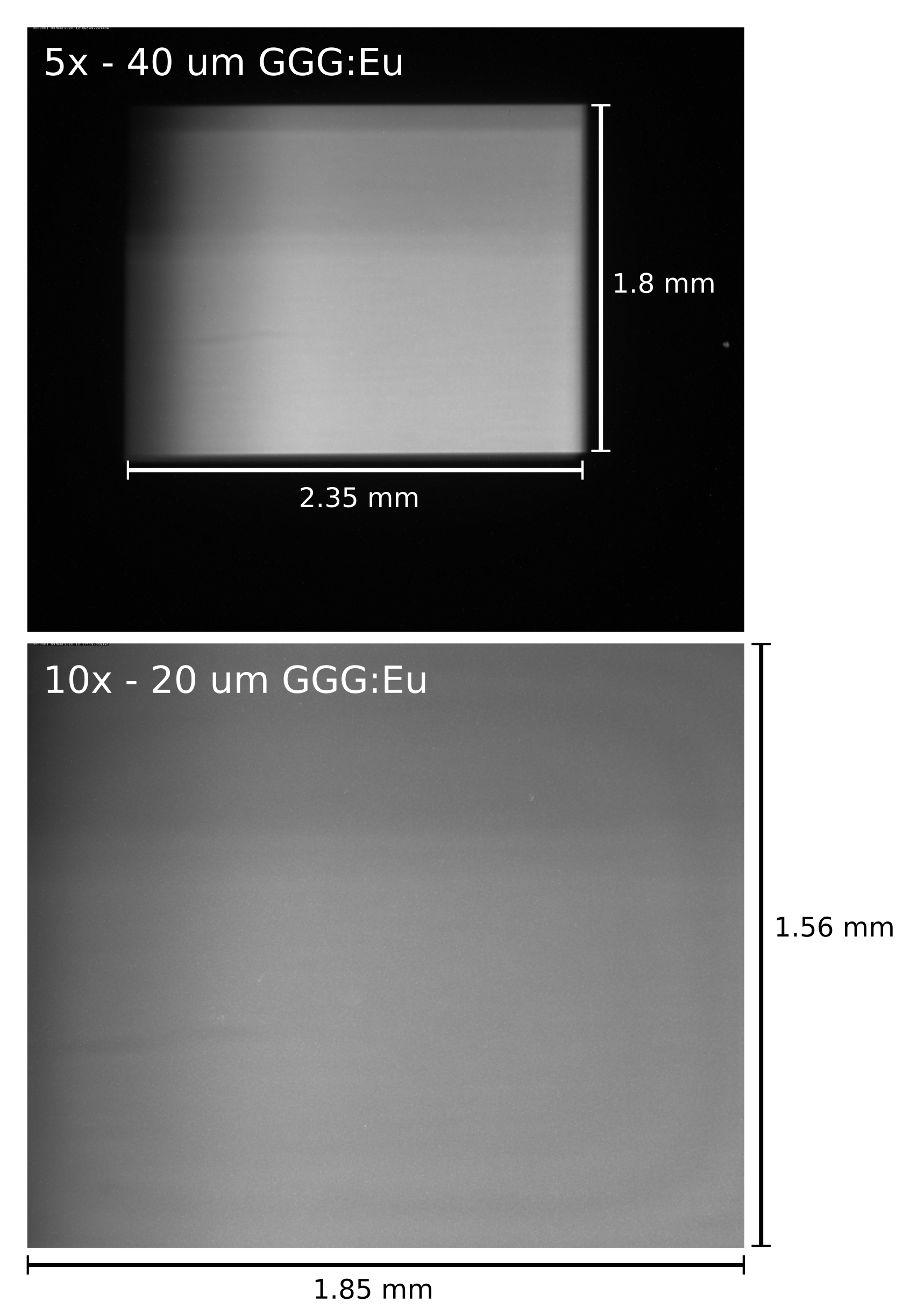
09-03-2020. Image frame rate tests (i.e. acquisition time tests). A number of images were captured at different acquisition times using the X-ray microscope equipped with a 5x and 10x objective and the PCO.edge 5.5 MP camera. The results can be found in this PDF.
We prefer to attenuate the X-ray beam with a 50 mm Al block before the scintillator to reduce the heat load to less than 1 W/mm2. Previously during a test, a 20 mm Al block was placed and the scintillator instantaneously broke by thermal fracture from direct beam exposure. Users are therefore encouraged to build an X-ray transparent assembly as much as possible. The main result of this test is that the weakly contrasting rock layers in a large cut assembly remain visible without significant image quality reduction down to 0.001 s exposure times, after simple image data treatment.
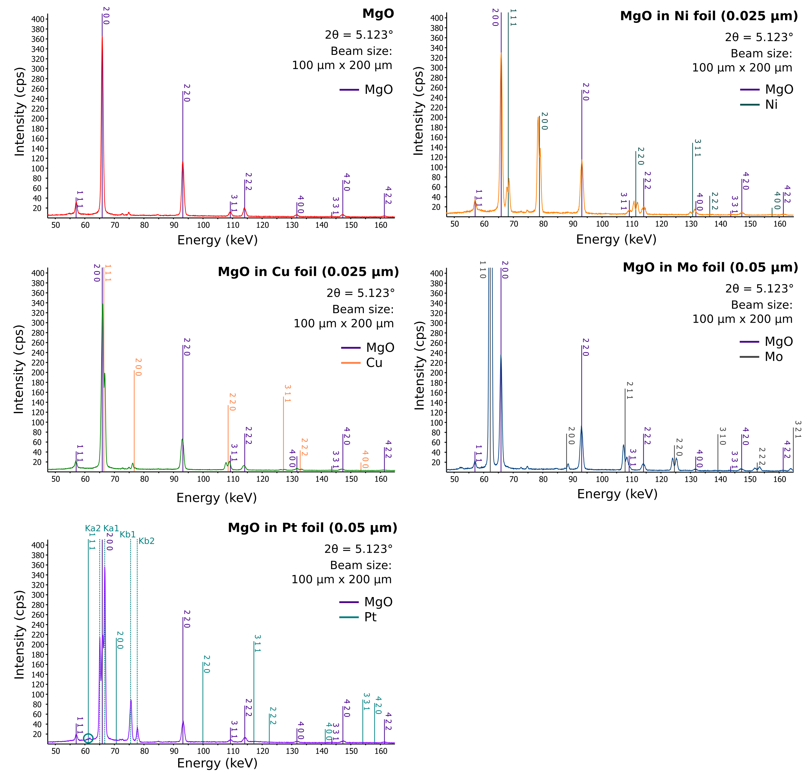
06-03-2020. Diffraction patterns obtained with MgO and MgO in various metal capsules (which did not melt or react). Note that except for Pt, all capsules, 2 mm in diameter, produce reflections indicating that the gauge volume for diffraction is larger than the capsule with MgO. This remains true even for a pencil beam of 50 um x 50 um. The collimator slit with horizontal gap of 50 um may be too large if a gauge volume smaller than 2 mm in the beam direction is required (at 2theta ~5°). In the case of Pt, the real problem are the fluorescence lines, which cannot be eliminated. The sample was surrounded by 14 mm of pyrophyllite to simulate beam attenuation in a cell assembly.
06-03-2020. MgO in thin Au foil (0.05 mm). The gold capsule immediately melted upon exposure to the X-ray beam. Therefore, temperature increased instantaneously to a value greater than 1060 °C! The sample was surrounded by 14 mm of pyrophyllite to simulate beam attenuation in a cell assembly.
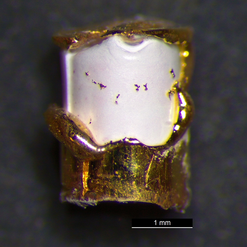
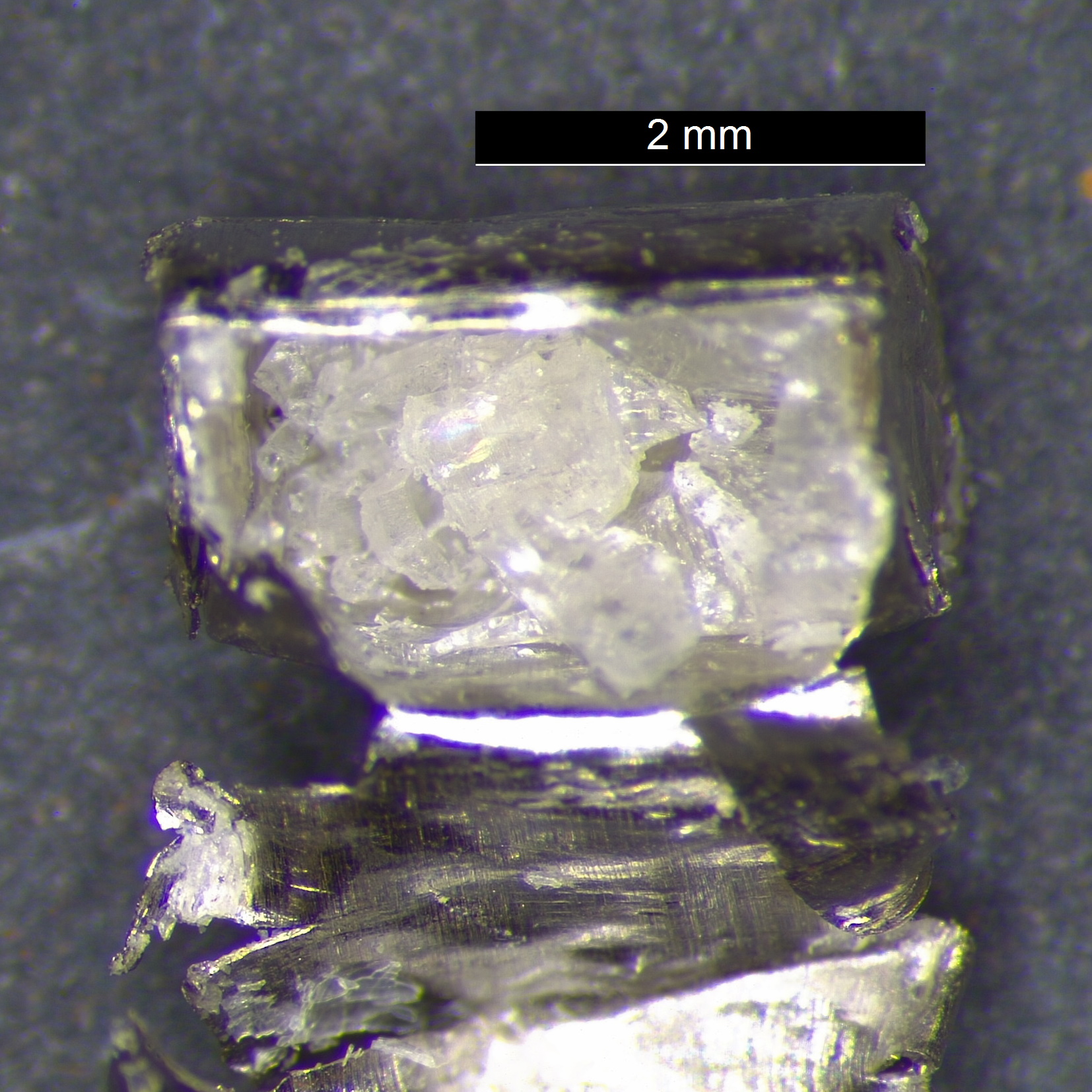
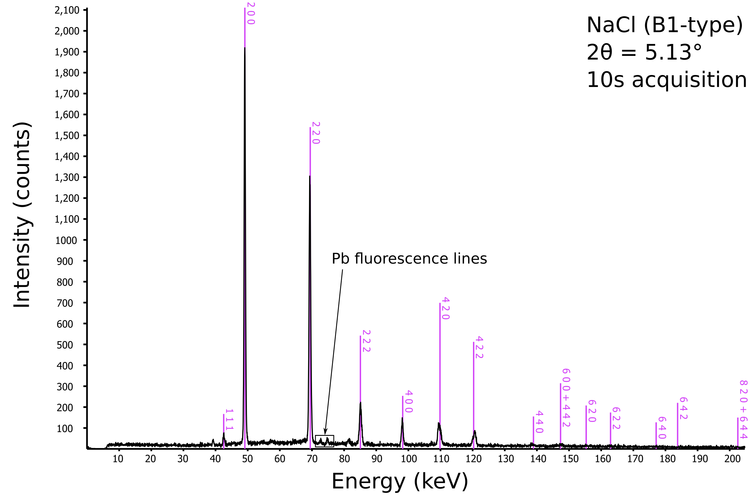
06-03-2020. Encapsulating NaCl in thin Pt foil (0.05 mm) or any other heavy metal is not a good idea for in situ X-ray diffraction at P61B. The white beam heats up Pt via X-ray absorption. The fairly X-ray transparent sample inside (NaCl) melted, this was directly observed using radiography on NaCl in a Ta capsule. This also would have happened with Pt, since the recovered sample is transformed from a powder to large quenched crystals. Therefore, a full beam exposure on a thin Pt or Ta foil (0.05 mm) will heat up the sample inside to over 800 °C! Another side effect is contamination of the fluorescence lines of metals in the diffraction pattern. The sample was surrounded by 14 mm of pyrophyllite to simulate beam attenuation in a cell assembly.
04-03-2020. We checked the diffraction conditions on NaCl. The diffracted beam size was 50 um (h) x 100 um (v). With the help of new Pb (lead) shielding on the Ge detector, the background and Pb fluorescence lines are nearly completely removed, resulting in a clean diffraction pattern.
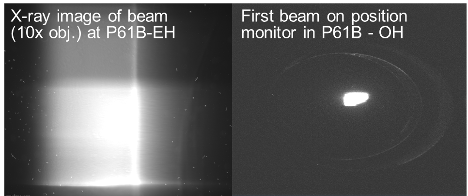
04-03-2020. After the reboot of the storage ring of PETRA III, the beam was checked again at P61B. The beam position has shifted and it appears a slightly larger beam is now available. The shadows are a result of upstream optics.
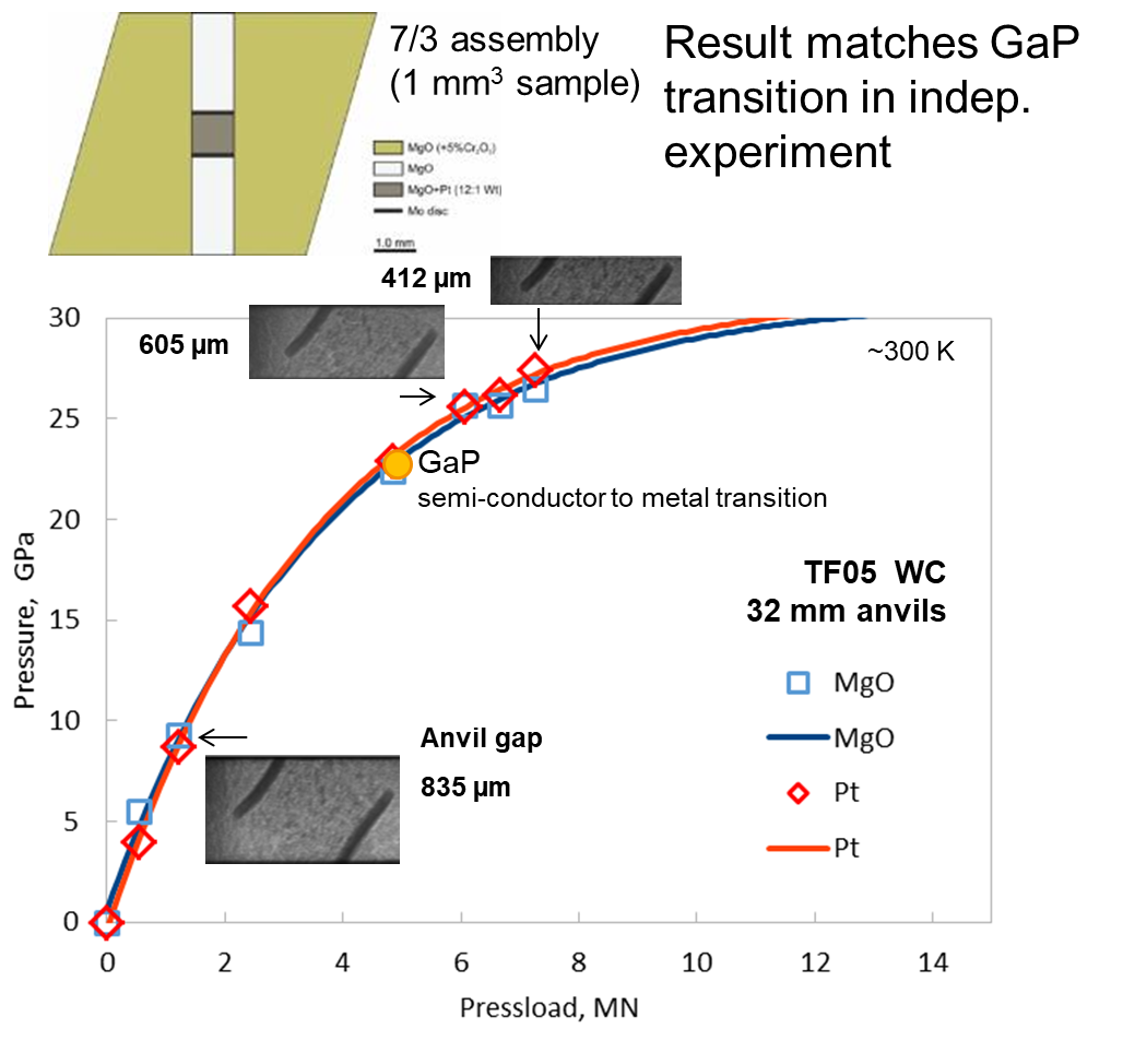
04-12-2019. New pressure calibration test with in situ X-ray diffraction on a mixture of MgO and Pt to determine the sample pressure up to 27 GPa at increasing press loads using the Equations of State of these materials. The ultra-hard WC anvils remain intact to date even after heating to 2000 K. This type of experiment using 7/3 assembly may be considered routine to 27 GPa or possibly to even higher pressures.
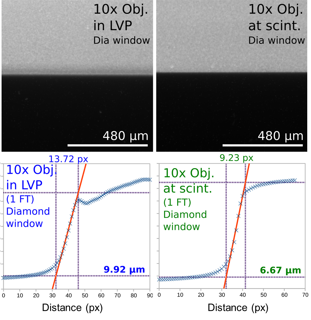
02-12-2019. An image resolution test was carried out using the 10x objective on the X-ray microscope. The edge of a small WC anvil was imaged at the scintillator and in the LVP (~3 m from the scintillator). We used a GGG:Eu 10 µm thick scintillator, 1 FrontEnd heat load filter and a 50 mm thick Al block to attenuate the power in the beam. Flux below 50 keV is also removed. The results show that the anvil edge is reasonably sharp, especially closest to the scintillator (< 10 px). Due to the nature of the white beam, there cannot be any further substantial improvement in image resolution. Optimal results (a 1-2 pixel edge) would need to be obtained elsewhere using a collimated monochromatic beam (with little divergence).
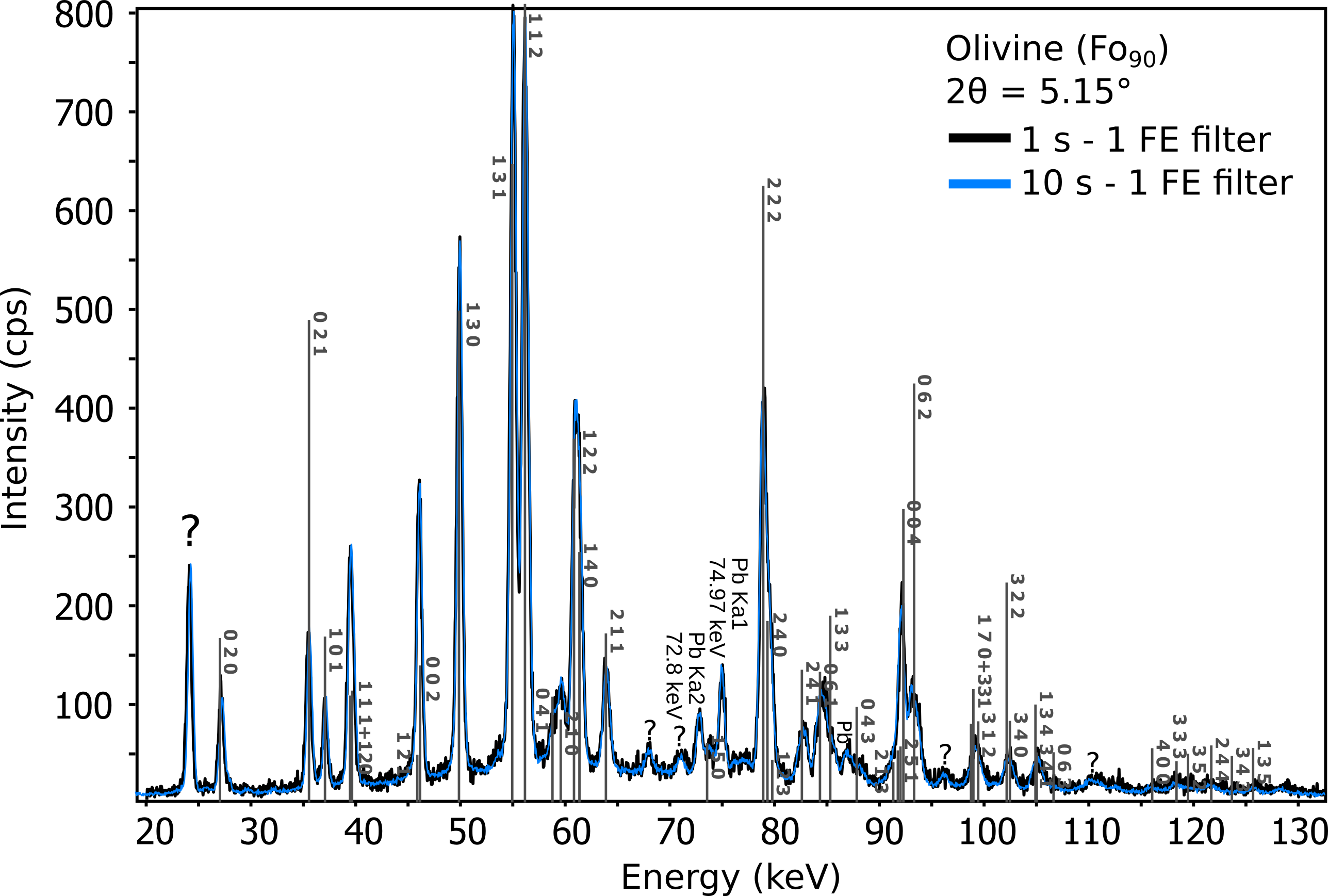
29-11-2019. ED-XRD was redone on a polycrystalline Fo90 olivine sample with fantastic results. A 10 s acquisition time is sufficient for publish-quality data. 1 s (or lower) acquisition times could be very interesting for fast reaction kinetics experiments that cannot be performed anywhere else in the world (phase transformation/nucleation processes at extreme conditions).
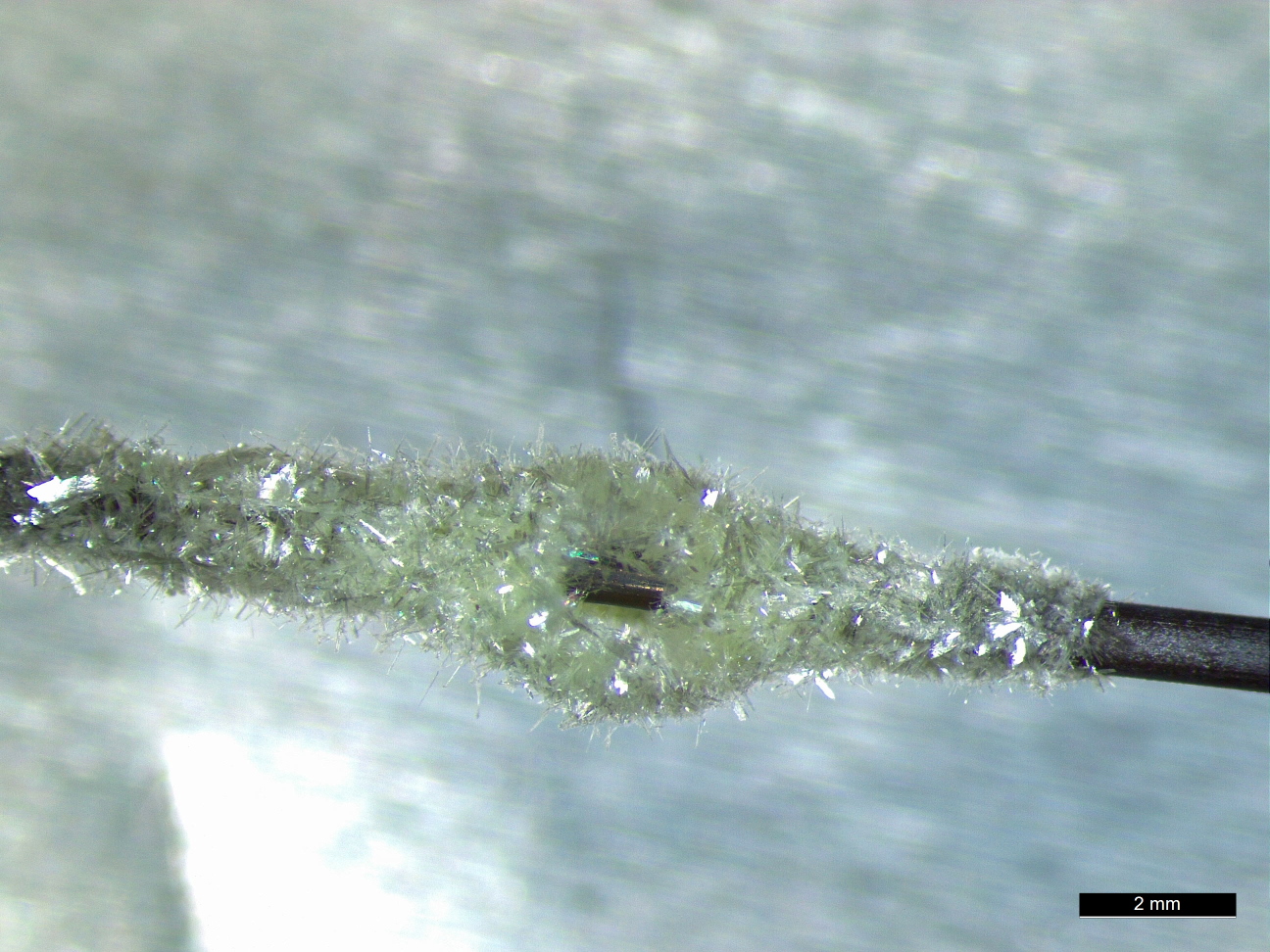
27-11-2019. The power in the beam is so high (even with 1 heat load filter inserted), that it's possible to oxidize everything, not just the air to ozone, but metallic objects such as Mo. A Mo rod was heated to at least 600 °C by the beam to initiate molybdite (MoO3) crystal growth within minutes of exposure.
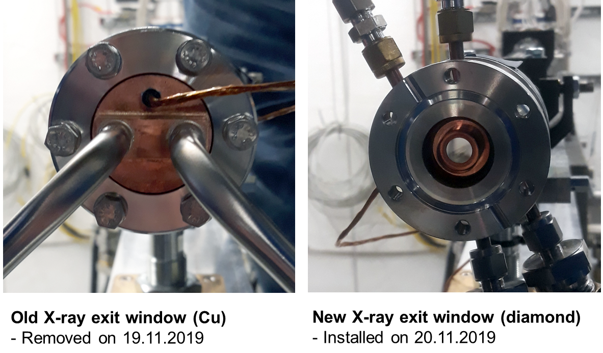
20-11-2019. Exit window replaced with CVD diamond for beam transport into the EH (P61B DESY). Full flux now obtainable (1 heat load filter must be used).
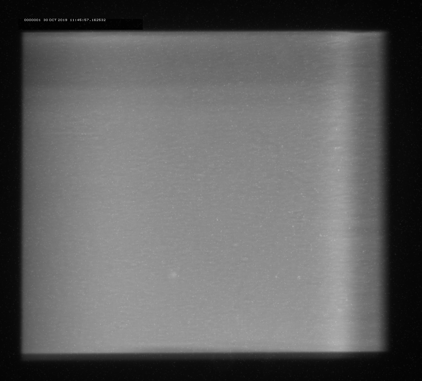
30-10-2019. Beam optics realignment for larger beam, approx. 2 mm (h) x 1.7 mm (v)
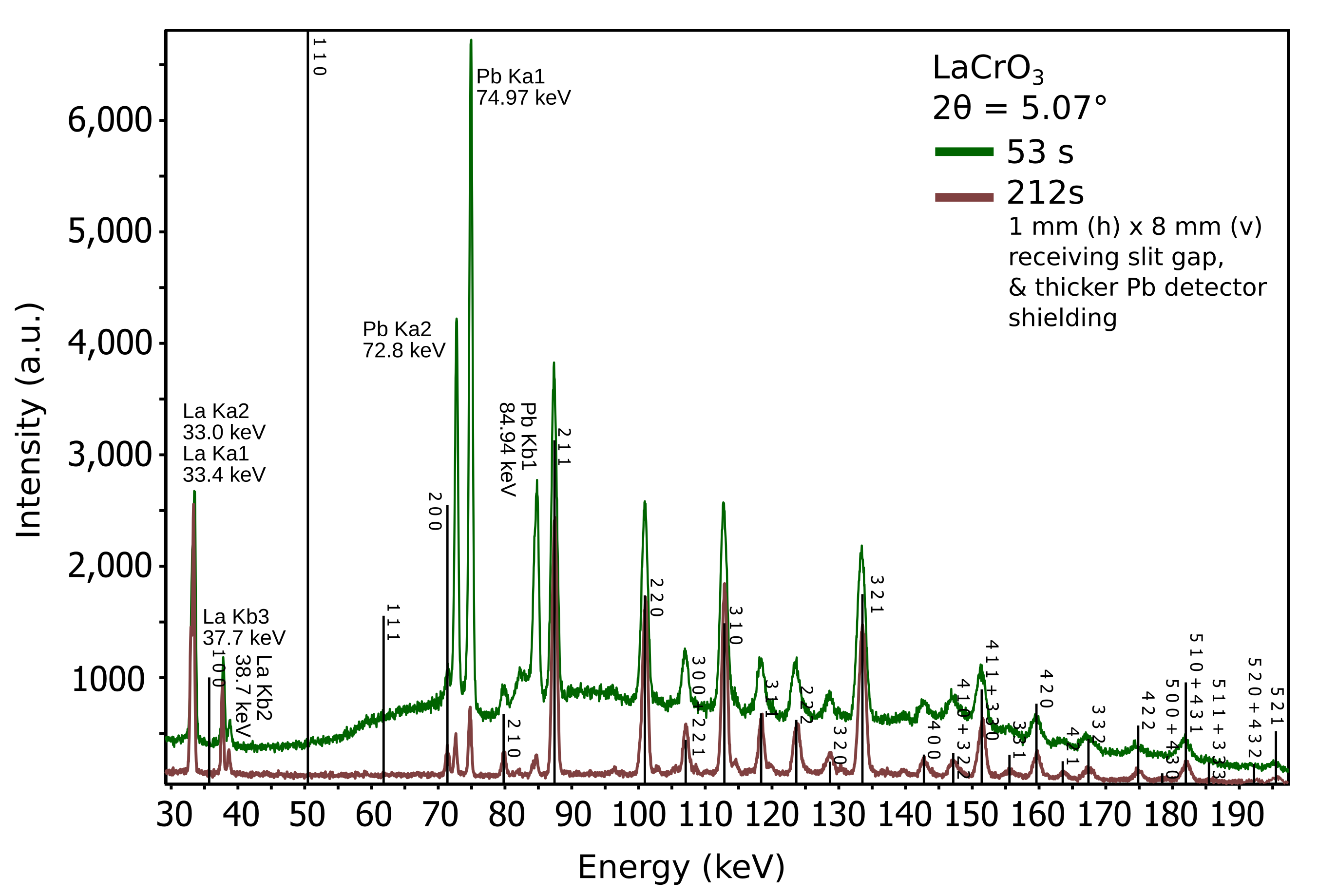
21-10-2019. Reduction of characteristic Pb X-ray emission from shielding and background reduction in diffraction pattern (e.g. LaCrO3).
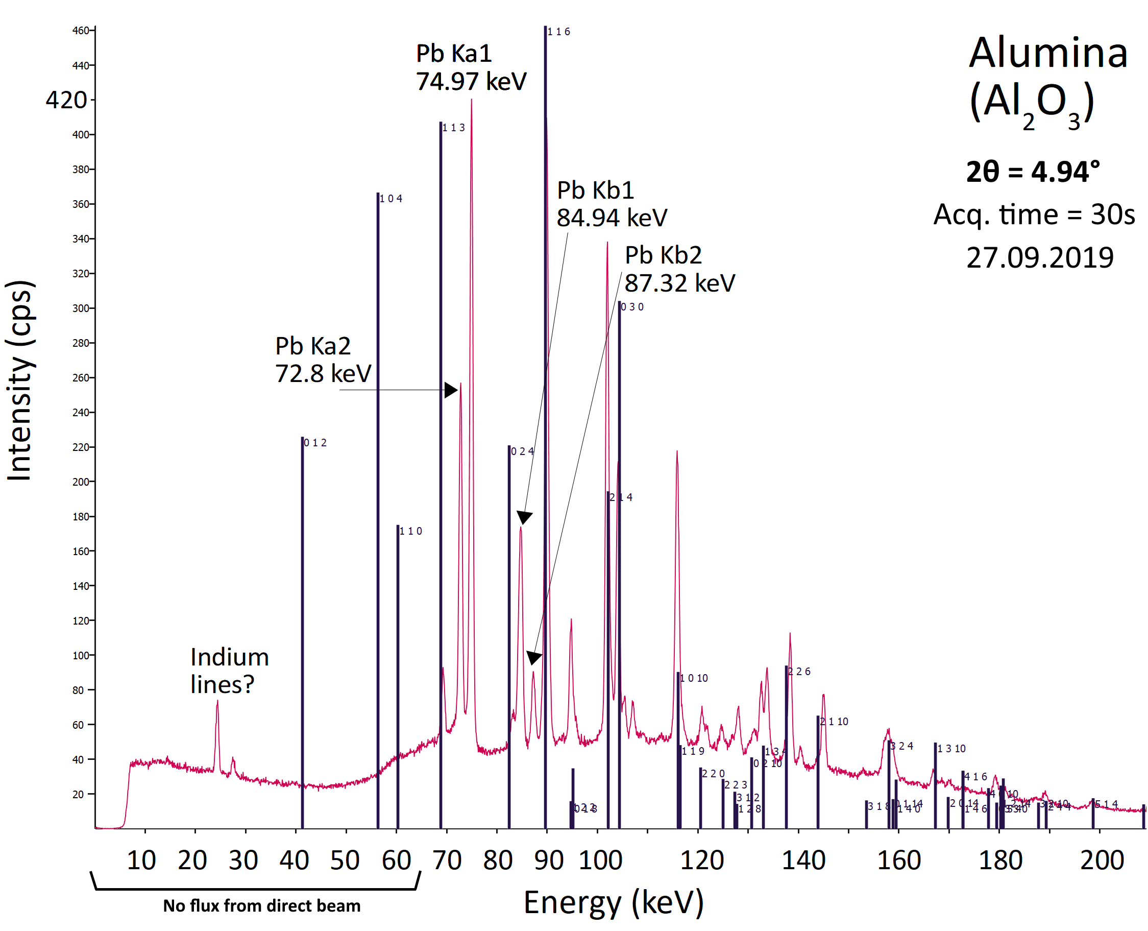
27-09-2019. First diffraction pattern obtained! Target sample was an Al2O3 rod.
23-09-2019. Addition of AC heating capability for the 6-ram LVP, courtesy of the Bayerisches Geoinstitut.
19-09-2019. Addition of a Ge-SSD (Germanium - solid state detector) on the temporary table. To be commissioned in week 39. (A 2nd Ge-SSD will not be added until May 2020 with the delivery of the detector pos. system from Kohzu)
23-08-2019. First deformation experiment using X-ray radiography.
Sample assembly: 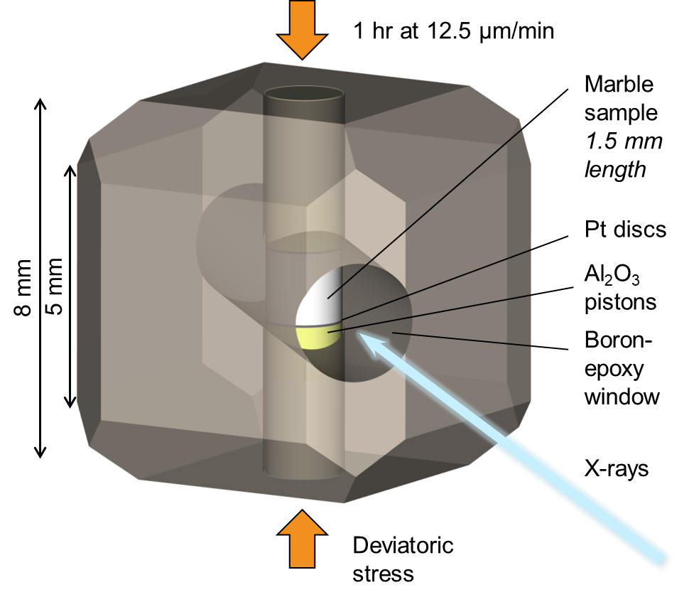
Deformation movie (gif) - Decompression movie (gif)
01-08-2019. First beam!
23-05-2019. The 6-ram LVP was upgraded this week with enhanced press modes:
1) Static compression
2) Deformation on the vertical rams
3) Uniaxial compression without the side rams.
For mode 2, multi-step deformation profiles for 2 independent rams moving at different or equal (and opposite) rates can now be constructed. The press is now part of the interlock system. Compression above 50 bar cannot continue while the hutch door is open (for safety reasons).
25-02-2019. Video! Go here to watch a run through the Paul P. Ewald hall of PETRA III, showing the construction status of P61 and P62.
15-02-2019. NEW! Leica M80 stereo microscope with HD (10 Mpix) camera now available. ● 6-ram LVP and press software to get major upgrade - 3 modes of compression, and more. ● Standard assemblies to be provided to users, published here. ● X-ray microscope system to be tested at P21.2 this February. ● Temporary detector positioning system design ready, construction starting soon. ● Twin Ge-SSD high count rate detector system arriving May 22, 2019. ● Complete detector positioning system eta. Nov 2019. ● First beam eta. now Summer 2019.
21-11-2018. Beamline construction update: Call for tender for cooling water, compressed air and air conditioning ends 13-12-2018. If successful, work is anticipated to begin at the end of January 2019 or early February 2019.
19-11-2018. The call for tender for the detector positioning system is advertised here.
15-11-2018. Regrettably, due to delays with a call for tender for the detector-collimator-slit positioning system, this system will not be ready until end of 2019. This will delay first high-P in situ experiments using X-rays. However, the detector and camera systems will be commissioned from early Summer 2019 using a temporary positioning solution. First beam is still expected for May 2019.
01-10-2018. The official 3rd LVP workshop was hosted on 01 and 02 October 2018. A good number of some 40 participants attended. For details please visit the archived workshop website here.
16-08-2018. First light in the newly completed Front End of P61! Have a look!
14-08-2018. Register now for the noon-to-noon workshop on focused on in situ extreme conditions research at the P61B LVP beamline using synchrotron X-rays. Visit the website here!
10-08-2018. P61B has a benchtop X-ray diffractometer with strip detector available for use. If interested, please check the webpage in the menu on the left.
14-05-2018. The LVP is reconnected and in use again. Please contact me with a project if you are interested. Bear in mind some calibration work is still ongoing.
25-01-2018. DESY User Meeting Xtreme Conditions session presentation: Status of P61B
18-01-2018. Some calculations of the beamline were done. See here.
20-12-2017. Important notice! The LVP is disconnected until the concrete hutches of P61 have been built. (Completed by end of March 2018).
04-11-2017. The LVP beamline P61B was showcased on DESY day (Nacht des Wissens).
16-10-2017. A 2 day Xtreme Conditions Workshop for PETRA IV took place.
01-06-2017. The Large Volume Press (LVP) is fully operational and users can apply for standalone use (see menu).
01-06-2017. The frontend and beamline are under construction and no X-rays are available. Estimated time of arrival of X-rays is projected for late 2018.