


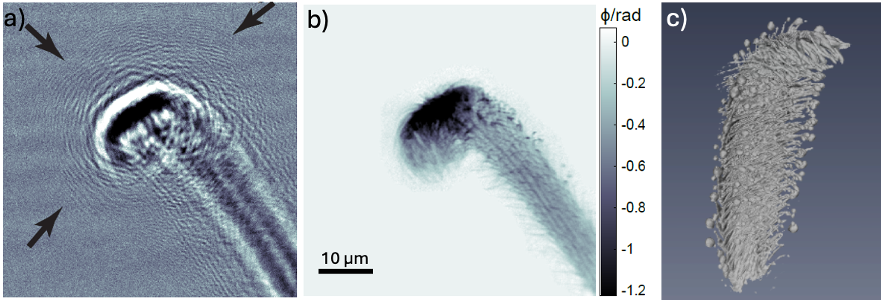
Transmission X-ray Microscopy. Siemens star test pattern in a) absorption and b) Zernike phase contrast, the structures of the third inner ring (48 nm - 37 nm) can clearly be resolved. c) Siemens star recorded with the dark-field method, developed at the beamline.

The P05 Nanotomography at PETRA III offers several imaging techniques for high-resolution, non-destructive analysis of materials and biological samples. These techniques allow for advanced imaging capabilities at the nanoscale, with applications ranging from material science to in situ studies. Each technique offers distinct advantages, and together, they cover a broad range of experimental needs, ensuring versatility for diverse research applications.
Transmission X-ray Microscopy (TXM)
The TXM at P05 offers absorption and Zernike phase contrast imaging, as well as the newly developed dark-field TXM method. Offering high-resolution imaging with fast scan times, it is particularly well-suited for in situ experiments across a diverse range of samples, even high-absorbing materials such as dense composites and metal-containing structures.
Its relatively large working distance of several centimeters allows for the integration of complex in situ environments, providing sufficient space for experimental setups without compromising imaging performance.
Key Features:
- Contrast: Absorption and Zernike phase contrast
- Resolution: Down to 40 nm
- Flexibility: FoV ranges from 30 µm to approximately 90 µm, accommodating small to larger samples (up to mm-sized tissue samples). Region-of-interest (ROI) scans are routinely possible.
- Fast Scans: Standard scans are performed in 15 min. Scan times range from 30 minutes for ROI scans to less than one minute for in situ applications.
- Energy Range: TXM operates between 8 – 17 keV, highest performance at 11 keV.
- XANES Imaging: Imaging at X-ray absorption edges has been successfully performed and is available upon request.
- Optics: Utilizes Fresnel Zone Plates (FZPs) and beam-shaping condensers with outermost zone widths of 50 nm for the highest resolution. 30 nm outer most zone width optics are available upon request (low flux).
Applications:
- Material Science: Detailed analysis of metals, composite materials, polymers, and other materials.
- Biological Studies: Imaging of radiation-resistant biological specimens at the nanoscale.
- Medicine: High-resolution imaging of large tissue samples for detailed structural analysis. ref
- Geology: Non-destructive imaging of rocks, minerals, and geological structures to study internal features.
- In Situ Experiments: Fast imaging allows for real-time radiography (50 ms time resolution) and 3D in situ indentation, such as material deformation.
Near-Field Holotomography (NFHT)
NFHT is a phase contrast imaging method that utilizes a cone beam configuration, providing exceptional flexibility for a wide range of applications. This technique is particularly well-suited for in situ imaging, allowing for real-time observation of dynamic processes under controlled conditions.
With a large working distance of several tens of centimeters, NFHT accommodates larger in situ environments, making it ideal for experiments requiring additional space. Additionally, its low X-ray dose enables imaging of sensitive samples in combination with water, ensuring minimal radiation-induced effects during experiments.
Key Features:
- Optics: Uses a Fresnel Zone Plate for focusing, with outermost zone widths of 50 nm, offering high resolution and smooth flat-field performance.
- Energy Range: Operates at 11 and 17 keV.
- Single Distance Phase Retrieval: Only a single distance is required for phase retrieval, making it ideal for in situ applications.
- Flexible FOV and Zooming: Allows for highly flexible field of view (FoV) from 30 µm to 2 mm, with zoom-in capabilities for ROI scans.
- Reduced Dose: Minimal X-ray dose to the sample, preserving its integrity during imaging.
Applications:
- Low-Absorbing Materials: High-contrast imaging of soft biological tissues, polymers, and porous structures.
- Biological Materials: Imaging of whole biological animals at the nanoscale, including radiation-sensitive samples.
- In Situ Experiments: Real-time imaging of processes involving water or other liquids, such as biological reactions or material behavior under changing environmental conditions.