


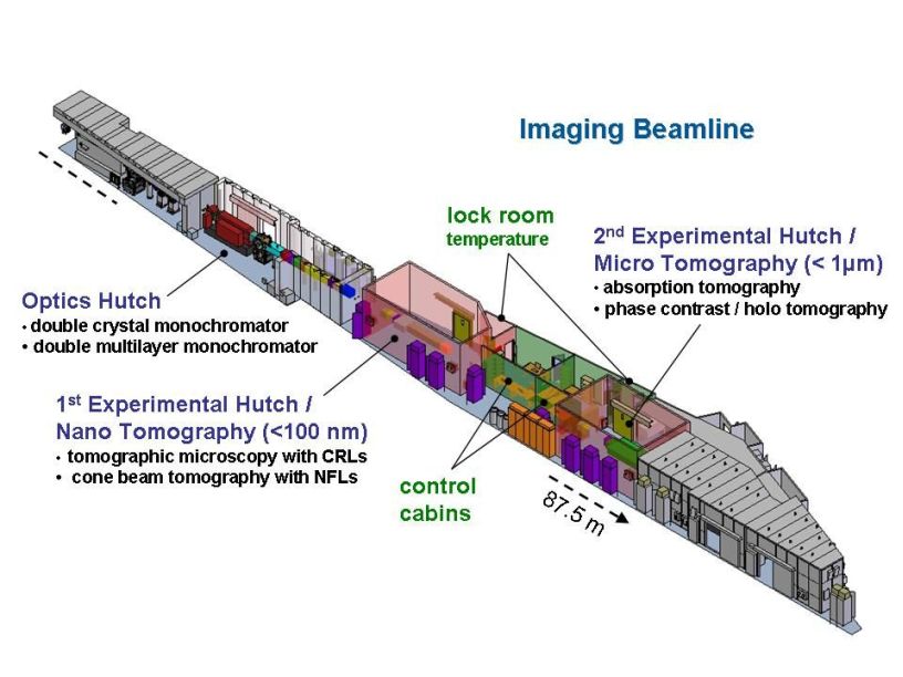
After the first workshop in September 2007 in order to optimize the optics and the setups for micro and nano tomography and to address the future user community, the current working design for the beamline (P05 in sector 4 of the future experimental hall) consists of a two meter undulator source, an optics hutch (OH) including two monochromators (DCM and DMM) and two independent experimental hutches EH1 and EH2 working alternatively.
|
The development of new materials highly demands the study of their three dimensional inner structures. Thus, in particular tomographic evaluation methods gain more and more importance in the materials characterization field. Characteristic length scales, which influence the structural properties, are often in the range of some micrometers down to a few nanometers.
Due to the extraordinary high brilliance of the new storage ring PETRA III, the extremely low emittance of 1nmrad and the high fraction of coherent photons even in the hard X-ray range an extremely intense and sharply focused X-ray light will be provided.
These unique beam characteristics will promote novel applications of tomographic techniques enabling ultra-fast in-situ measurements as well as highest spatial and density resolution. Additionally the highly coherent beam enables the application of phase contrast methods in an exceptional way.
Therefore, the HZG Research Centre Geesthacht takes active part in the PETRA III project by construction, operation and funding of the Imaging Beamline (IBL). This beamline will be optimized for micro and nano tomography applications.
Optics Hutch
To generate monochromatic X-rays two types of monochromators will be installed in the optics hutch. A silicon single crystal monochromator (DCM), designed by DESY will be used for tomographic methods, which need a very high monochromatization (e.g. vector tomography, absorption edge tomography, diffraction tomography). For applications, which need particularly high flux (e.g. for fast in situ experiments) a double multilayer monochromator will be designed. The energy range for both types of monochromators will be tunable between 5 and 50 keV.
Micro Tomography
Due to the divergence of the low-β X-ray beam in large distance from the source the field of view will be large enough to investigate samples of some millimetres diameter in (sub)-micrometer resolution.
To achieve spatial resolutions near the physical limit, high precision air bearing stages will be used for the sample rotation as well as for the camera movement. In addition a special sample positioning system was designed, which guarantee extremely low tilt errors of the samples.
Fields of application:
- Materials science (e.g. imaging and quantitative analysis of pores, cracks, precipitations, grain structures, small components, phase transitions an morphological transitions (in situ))
- Biology and geology (e.g. plants, insects, stones, soil)
- Medicine (e.g. implants, structures of bones, tissues, teeth)
Further a motorized microscope optics made of X-ray resistant glasses and an automatic sample changer will be implemented.
Nano Tomography
The feasibility to focus the X-ray beam on nanometer scale enables nano tomographic imaging. Two different dedicated nano tomography setups are planned. One of them realized by compound parabolic refractive lenses, the other using crossed cylindrical nano focus lenses, both to achieve a beam geometry for magnified imaging. In both cases a spatial resolution below 100 nm will be expected.
After completion of the Imaging Beamline IBL and the High Energy Materials Science Beamline HEMS at PETRA III four complementary tomography stations will be managed by the HZG at DESY. The HARWI setup and the BW2 setup are characterized by a large field of view and an excellent absorption contrast. The tomography facilities at PETRA III distinguish themselves with high flux at small fields of view and high coherence, i.e. they fulfil excellently the qualifications for phase contrast or holo tomography, for nano tomography and for high speed or in situ tomography.
|
DORIS III |
DORIS III |
PETRA III |
PETRA III |
|
|---|---|---|---|---|
|
HARWI |
BW2 |
Micro and Nano Tomography (IBL) |
High Energy Materials Science (HEMS) |
|
|
X-ray energies |
16 - 250 keV |
7 - 24 keV |
5 - 50 keV |
50 - 300 keV |
|
field of view |
up to 70 x 8 mm |
up to 20 x 4 mm |
up to 5 x 0.9 mm |
up to 2.4 x 0.5 mm |
|
coherence |
no |
no |
yes |
yes |
|
max. spatial resolution |
3 µm |
2 µm |
< 1 µm / < 100nm |
< 1 µm |
| Useful links |
|