


")
")
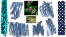
Amyloids can adapt their architecture depending on purpose and environment, revealed by a unique combination of techniques. Detail of the figure below (Credit: Authors/DESY)
")
")
What do the Blue Mountains tree frog and Alzheimer’s patients have in common? Not much at first sight – but there is one special form of protein structure that plays a crucial role for both. These protein fibres, called amyloids, are found in the brains of people suffering from the neurodegenerative Alzheimer’s disease. They are also found on the skins of amphibians (like the tree frog), where they play a very different and much more positive role: They destroy harmful germs. Meytal Landau, structural biologist at the Centre for Structural and Systems Biology CSSB, and her team are trying to find out whether the positive aspects of the one can be used in favour of the other.
Using a combination of techniques unique to DESY – X-ray crystallography at the PETRA III beamline P14 operated by EMBL Hamburg, the CSSB’s cryo-electron and light microscopes and DESY’s computing facilities – the scientists looked at the peptide citropin 1.3 derived from the frog. They used also other facilities and found that citropin 1.3 can form various shapes, from classic amyloid fibres, which are like tightly packed b-sheets stacked up, to α-helical assemblies and even multi-layered nanotubes with a unique mixed architecture. The atomic-resolution images show the different forms in very high detail, paving the way to a much better understanding of the unique properties of these peptides. The combination of techniques is key as well: While the X-ray crystallography revealed a wealth of amazing formations, the amyloid form was only seen by the cryo-electron microscopes.
The team thinks that the peptide’s impressive shape-shifting versatility comes from the necessity for amphibians to adapt to quickly changing and extreme environments – from hot to cold, wet to dry and mostly very dirty and full of microbes. They also found that citropin 1.3 interacts dynamically with cell membranes and forms tiny liquid droplets inside cells which effectively gives them the power to kill cells and perhaps to manipulate the genetic material.
“These findings reshape our understanding of amyloids as disease agents. They turn out to be versatile biological tools with specific functions and controls, beyond that of the DNA sequence level,” summarises Meytal Landau. “The next step would be to check how we can turn the versatility into a benefit for us, for example for developing antimicrobial strategies or smart biomaterials.”
In Alzheimer’s or Parkinson’s patients, the amyloid fibres form plaques that interfere with normal cell function. The question is: Why are they there in the first place? Can humans form amyloids to fight pathogens, and could these diseases be signs of successfully averted brain infections in a younger age? Can the build-up of plaques be prevented when there’s more awareness of them earlier on, and can we in future make our own antimicrobial agents and materials that respond to environmental changes? “The field is buzzing at the moment,” says Landau. Her team’s results are set to make advancements possible.
The studies weren’t easy, though. “The biggest challenge was capturing the extreme structural diversity of citropin 1.3 and its strong sensitivity to environmental conditions”, Landau explains. The peptide formed multiple fibril types simultaneously, which is why the team needed to use different methods to capture them while knowing that there are even more. “Another major hurdle was combining structural, biophysical and cellular data because the peptide’s activity changes with its shape, and its shape shifts over time and in response to interactions with membranes and intracellular components.” With current technologies, it is essentially impossible to capture precisely the dynamic structural changes the peptide undergoes in its natural environment. Overcoming these limitations will be a key challenge for next-generation imaging and structural biology technologies in the years ahead – including future lightsources like PETRA IV.
(Partly from DESY News)
Reference:
F. Strati, M. P. Cali, Y. Bloch, S. Mostafavi, J. Monistrol, A. Golubev, B. Rayan, E. Gustavsson, M. Landau, “ Structural and Functional Versatility of the Amyloidogenic Non-Amidated Variant of the Antimicrobial Peptide Citropin 1.3.” Adv. Sci. (2025) DOI: 10.1002/advs.202503997