


")
")
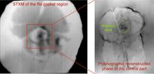
Two views of an extreme-states experiment: To the left is an X-ray micrograph of the sample set up, which consisted of a piece of elemental iron surrounded by solid oxygen, itself surrounded by a rhenium gasket within a diamond anvil cell creating intense pressure. To the right is a ptychographic reconstruction of the area of the sample hit by X-rays, shown with a green circle. In that area using their new ptychographic method, the team could reconstruct the oxidation of the iron being melted by the intense pressure. An extreme-states experiment of this kind has not before been imaged in this way. (Credit: Tang Li, DESY)
Sometimes a change of perspective can make a world of difference. A team of scientists from DESY and MAX IV as well as University of Bayreuth has rearranged the method in which one can use an X-ray beam to image a sample without using high-quality lenses. The method, called ptychography, has been widely used at synchrotrons and free-electron lasers to analyse the inner workings of materials quickly enough while avoiding major damage to the sample by the X-rays. The team has turned the standard method of ptychography on its head: Instead of moving the sample around the X-ray beam, they have figured out how to move the X-ray beam itself in a way that does not alter the properties of the X-rays while still accomplishing the effect of ptychographic analysis. Moreover, they have tested the method on a sample that is in and of itself difficult to move – short-lived states of matter under extreme conditions of pressure and temperature. The team has published their findings in the Proceedings of the U.S. National Academy of Sciences (PNAS).
X-ray ptychography has become, in recent years, a standard technique in the toolbox of researchers using X-ray light sources. In a wide variety of fields, including biology and geology, the technique has been critical for imaging the interiors of samples up to atomic-scale detail non-destructively, revealing details on a scale that methods of light and electron microscopy cannot reach. Up to now, ptychography has been accomplished by using extremely precise sample movers that would change the position of the sample relative to the X-ray beam by tiny lengths – sometimes to the nanometre level – creating a grid pattern of sequentially imaged spots that eventually revealed the full image. Called high-resolution phase-contrast imaging, it has provided insights into the nanoscale structures of tiny biological structures, mineral deposits, computer chips and much more.
However, it has been difficult to perform ptychography on certain kinds of samples. For example, samples being compressed to high pressures cannot easily be moved in the required way, partly because devices used to generate such pressure are too heavy to move so precisely in the required way and too bulky, requiring a very large optics–sample distance. The team involved in this research tried an alternative method of ptychography as a way to address this issue.
In a technique developed and implemented at the P06 beamline at PETRA III, the team inserted a high-precision fast-oscillating mirror to move the X-ray beam across the sample and developed a new data interpretation approach. The mirror is precise enough to replicate the motions of a movable sample frame or stage, allowing the beam to scan the sample and produce similar results.
The team tried this on extreme pressure studies using a common device to generate extreme pressures: a diamond anvil cell, wherein a material is compressed to a high degree between two diamonds. As a first application, they compressed a sample of iron surrounded by solid oxygen, irradiated it with a laser beam and performed ptychography on the resulting bulge of molten metal created by the laser heat under high pressures, analysing the oxidation of the metal at high temperatures at the nanoscale as a proof-of-concept.
“The context of this research is about making imaging in extreme environments from impossible to possible,” says Tang Li, a DESY researcher who is first author of the paper. “By steering the X-ray beam instead of translating the sample, we visualised iron oxidation at 50 gigapascals with nanometre resolution, expanding the reach of X-ray nanoimaging to extreme environments and opening the possibility of in operando studies in bulky and heavy sample environments.”
This adaptation of ptychography opens many more avenues for studies that will be performed at DESY’s future X-ray light source, PETRA IV. When combined with the enhanced spectral brightness of a fourth-generation synchrotron beam such as that from PETRA IV, the beam-scanning implemantation of ptychography will enable investigations of materials that have short lifetimes, that are exceptionally sensitive to motion or that are too heavy or reactive to move – delivering nanoscale studies of materials in extreme states.
The research team comprised scientists from DESY (PETRA III and CXNS), the University of Bayreuth (Germany) and the MAX IV Synchrotron in Lund (Sweden).
(Partly from DESY News)
Reference:
Li et al., "High-Resolution Ptychographic Nanoimaging under High Pressure with X-ray Beam Scanning", PNAS (2025), DOI: 10.1073/pnas.2514163122; DESY PubDB: https://bib-pubdb1.desy.de/record/618929/