


")
")
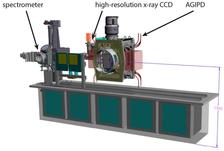
Detector bench: Flexible platform to support various X-ray detectors (Image: DESY).
")
")
")
")
")
")
Workpackage 1: A. Schropp (DESY)
Within the HIBEF-consortium we aim to develop parts of the X-ray instrumentation at the HED-instrument required to optimally probe matter in extreme conditions with the XFEL beam. Main parts of this workpackage are a movable detector bench supporting a suite of detectors to collect the various X-ray scattering or imaging signals as well as a focusing setup to produce tightly focused beams.
Detector Bench
Detectors at the end of the HED hutch will be supported by a bench, which is movable along the XFEL beam. It is designed to support various detectors required for X-ray diffraction, spectrometry, magnified phase-contrast imaging (PCI), or small-angle X-ray scattering (SAXS). An 'Adaptive Gain Integrating Pixel Detector' (AGIPD) will be implemented as a main detector for X-ray scattering, which can be operated at the full frame rate of 4.5 MHz of the European XFEL.
Focusing Setup
The European XFEL will provide a X-ray beam with outstanding properties of high spectral peak brightness, short and tunable wavelength, short pulse duration and narrow bandwidth. This beam is especially suitable for time-resolved studies. However, fast processes in matter involve short length scales so that meaningful measurements often require high resolution both spatially and temporally. Thus, XFELs with intense femtosecond scale pulse durations necessitate the development of high resolution imaging techniques, which require magnified imaging in real space or diffractive imaging in reciprocal space. In either case, the highest resolution will require focusing of the X-ray beam.
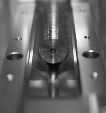
For this reason, a nano-focusing setup will be implemented at HED to strongly focus the XFEL beam to a size of about 100nm (FWHM) and even smaller. Current developments are based on a prototype setup, which was built for previous experiments at the MEC-instrument of the Linac Coherent Light Source (LCLS, US). It will be adapted to the experimental environment at the HED-instrument and technical components will be transferred to the European XFEL during the beginning of 2018. It will allow to install multiple X-ray optics and will provide all required degrees of freedom to align sample and optics.
The X-ray focusing will be based on a stack of compound refractive X-ray lenses made of beryllium (Be-CRLs). Multiple of these lenses need to be stacked to achieve the tight focusing conditions.
Magnified Phase-Contrast Imaging
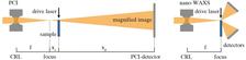
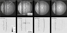
With this nano-focusing setup we also aim to develop X-ray imaging techniques at the European XFEL. A suitable method is for example magnified X-ray phase-contrast imaging, which can be used to capture snapshots of fast processes with high spatial resolution.
A diamond sample is imaged in a magnified projection geometry as schematically shown (see figures). The phase-contrast image is obtained by focusing the XFEL beam closely in front of sample and detecting the transmitted wave field further downstream. Alternatively, the sample can be moved into the focal plane of optics to determine the localatomic structure of a sample using wide-angle x-ray scattering (WAXS).