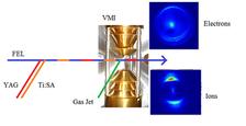
Experimental Setup for FEL-based Time-Resolved Photoelectron Diffraction Experiments. Free-Electron Laser (FEL), YAG, and Ti:Sa laser beams are focused on the interaction point in a velocity map image (VMI) spectrometer, where they interact with the target molecules delivered from a cold molecular beam. The molecules are adiabatically laser-aligned by the YAG laser, a photoreaction is triggered by a pulse from the femtosecond Ti:Sapphire laser, and the evolving molecules are probed by the FEL for different delays between Ti:Sapphire laser and FEL. Photoelectrons and fragment ions are detected on two microchannel plate (MCP) detectors equipped with a phosphor screen which is read out shot-by-shot with a CCD camera.
Photoelectron diffraction is a well-established method in solid state and surface physics, in which angle-dependent scattering of inner-shell photoelectrons yields information about the environment of the electron emitter. It is used, for example, to determine orientation and geometry of molecules adsorbed on surfaces [see, e.g., Woodruff1994, Fadley2008], thus enabling a detailed insight into physical or chemical processes such as catalytic reactions. In principle, photoelectron diffraction can also be used to study the structure of free molecules in the gas phase. However, this is hampered by the typically random orientation of gas-phase molecules, which averages out most of the intensity variations that contain the structural information. An experimental break-through was the development of angle-resolved photoelectron-photoion coincidence techniques in the 1990s [Cherepkov1992, Shigemasa1995, Heiser1997], which enabled the determination of molecular-frame photoelectron angular distributions (MFPADs) and gave access to an unprecedented level of detailed information [Gessner2002, Motoki2002, Schoeffler2008, Rolles2005, Akoury2007, Zimmermann2008] (see also Photoelectron-Photoion Coincidence Experiments with Synchrotron Radiation). When interpreting MFPADs in terms of photoelectron diffraction [Landers2001, Rolles2006, Zimmermann2008], direct information on the geometric and electronic structure of the molecule can be obtained, e.g. by comparing the measured diffraction patterns and molecular-frame photoelectron distributions to multiple scattering calculations [Dill1976, Muino2002, Golovin2005].
The determination of MFPADs via photoelectron-photoion coincidence requires light sources with a very high repetition rate since the coincidence are built up as a sum of many hundreds of thousands of individual events. An alternative method, which allows measuring MFPADs of an ensemble of aligned molecules and which is thus better suited for lower repetition light sources such as the currently operating Free-Electron Lasers, is to actively align the molecules in the sample by adiabatic or impulsive alignment in a strong laser field [Stapelfeldt2003]. Given the short pulses provided by Free-Electron Lasers with pulse length on the order of a few tens of femtoseconds or below, this allows performing femtosecond time-resolved photoelectron diffraction experiments on gas-phase molecules, where a photoreaction is triggered by a short pump pulse from an IR or UV laser and the evolution of the molecular system is probed with atomic spatial and femtosecond temporal resolution by the Free-Electron Laser [Boll2013].
A schematic of our setup used for FEL-based time-resolved photoelectron diffraction experiments is shown in Figure 1. To date, we have performed several time-resolved experiments both at LCLS and FLASH on a variety of molecules such as carbonyl sulfide (OCS), dibromobenzene, methyl iodide, p-fluorophenylacetylene (p-FAB) and others, and further experiments are planned.
Own References:
D. Rolles et al., Femtosecond X-Ray Photoelectron Diffraction on Gas-Phase Dibromobenzene Molecules, J. Phys. B 47, 124035 (2014).
R. Boll et al., Imaging Molecular Structure through Femtosecond Photoelectron Diffraction on Aligned and Oriented Gas-Phase Molecules, Faraday Discussions 171 (in press).
R. Boll et al., Femtosecond Photoelectron Diffraction on Laser-Aligned Molecules: Towards Time-Resolved Imaging of Molecular Structure, Phys. Rev. A 88, 061402(R) (2013).
D. Rolles, Trendbericht Physikalische Chemie 2012: Molekülkino – Experimente mit Freie-Elektronen-Lasern, Nachr. a. d. Chemie 61, 313 (2013).
D. Rolles et al., Isotope-induced Partial Localization of Core Electrons in the Homonuclear Molecule N2, Nature 437, 711-715 (2005).
B. Zimmermann, D. Rolles, B. Langer, R. Hentges, M. Braune, S. Cvejanović, O. Geßner, F. Heiser, S. Korica, T. Lischke, A. Reinköster, J. Viefhaus, R. Dörner, V. McKoy, and U. Becker, Coherence and Coherence Transfer in Molecular Photoelectron Double-slit Experiments, Nature Phys. 4, 649-655 (2008).
R. Díez Muiño, D. Rolles et al., Angular Distributions of Electrons Photoemitted from Core Levels of Oriented Diatomic Molecules: Multiple Scattering Theory in Non-Spherical Potentials, J. Phys. B 35, L359-L365 (2002).
Other relevant publications in the research field:
D. P. Woodruff and A.M. Bradshaw, Adsorbate structure determination on surfaces using photoelectron diffraction, Rep. Prog. Phys. 57, 1029 (1994).
C. S. Fadley, Atomic-level characterization of materials with core- and valence-level photoemission: basic phenomena and future directions, Surf. Interface Anal. 40, 1579 (2008).
N.A.Cherepkov, A.V.Golovin, and V.V.Kuznetsov, Photoionization of oriented molecules in a gas phase, Z. Phys. D, 24, 371 (1992).
E. Shigemasa et al., Angular Distributions of 1ss Photoelectrons from Fixed-in-Space N2 Molecules, Phys. Rev. Lett. 74, 359 (1995).
F. Heiser et al., Demonstration of Strong Forward-Backward Asymmetry in the C1s Photoelectron Angular Distribution from Oriented CO Molecules , Phys. Rev. Lett. 79, 2435 (1997).
O. Geßner et al., 4s-1 Inner Valence Photoionization Dynamics of NO Derived from Photoelectron-Photoion Angular Correlations, Phys. Rev. Lett. 88, 193002 (2002).
S. Motoki et al., Direct Probe of the Shape Resonance Mechanism in 2sg-Shell Photoionization of the N2 Molecule, Phys. Rev. Lett. 88, 063003 (2002).
D. Akoury et al., The Simplest Double Slit: Interference and Entanglement in Double Photoionization of H2, Science 318, 949 (2007).
M. Schöffler et al., Ultrafast Probing of Core Hole Localization in N2, Science 320, 929 (2008).
D. Dill, J. Siegel, and J.L. Dehmer, Spectral variations of fixed-molecule photoelectron angular distributions, J. Chem. Phys. 65, 3158 (1976).
A.V. Golovin et al., Inner-shell photoelectron angular distributions from fixed-in-space OCS molecules, J. Phys. B 38, L63 (2005).
A.V. Golovin et al., Inner-shell photoelectron angular distributions from fixed-in-space OCS molecules: comparison between experiment and theory, J. Phys. B 38, 3755 (2005).
H. Stapelfeldt and T. Seideman, Aligning molecules with strong laser pulses, Rev. Mod. Phys. 75, 543 (2003).


