


")
")
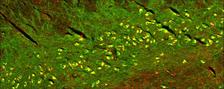
Iron deposits (red) in brain tissue: Using X-ray fluorescence measurements at DESY's X-ray light source PETRA III, researchers were able to map the iron concentrations in nerve cells of the substantia nigra (region in the brain stem). The cell bodies (yellow) of the dopamine-producing nerve cells have a very high iron concentration. (Image Credit: E. Kirilina, Department of Neurophysics, MPI for Human Cognitive and Brain Sciences, Leipzig, Germany)
The neurotransmitter dopamine is primarily known as the happiness hormone that controls our motivation in the brain's reward system. However, the neurotransmitter also acts as lubricating oil for our fine motor skills and regulates the movements of our muscles. If dopamine-producing nerve cells die off, affected people experience movement disorders such as tremors or muscle stiffness. The diagnosis: Parkinson's disease. Researchers suspect that the reason for the death of nerve cells is excessive iron concentrations in the brain.
A team of researchers from Germany and the UK has now developed a method that can be used to determine the iron concentration in the affected regions. With the participation of DESY researchers Gerald Falkenberg and Dennis Brückner, the team led by Evgeniya Kirilina from the Max Planck Institute for Human Cognitive and Brain Sciences in Leipzig, was able to determine possible toxic iron concentrations from MRI (magnetic resonance imaging) measurements of cells using DESY's brilliant X-ray light source PETRA III. The work could contribute to the development of early diagnoses for Parkinson's disease.
Parkinson's disease is one of the most common diseases of the nervous system, affecting around 200,000 people in Germany alone. There is currently no cure for the disease. The typical Parkinson's symptoms are caused by damaged nerve cells in the substantia nigra, an area in the brain stem. Damaged or dead nerve cells no longer produce enough dopamine or any dopamine at all - the lack of dopamine disrupts signal transmission between the nerve cells.
Iron is required for dopamine production in the nerve cells, and the corresponding nerve cells in the substantia nigra are therefore susceptible to both iron deficiency and excessive amounts of iron. Too much intracellular iron can be toxic, leading to the degeneration and death of neurons in the substantia nigra. "Oxidative stress caused by iron is considered a possible cause of the death of dopamine-producing nerve cells," says DESY researcher Gerald Falkenberg, head of beamline P06 at DESY's research X-ray source PETRA III. "That is why we have been looking for methods to measure the amount and distribution of iron in the brain over the course of a person's life." According to Falkenberg, this should also be possible for patients in hospitals in the future.
Researchers do not yet know what levels of iron are pathological for humans, and the course of cellular iron levels over the course of a person's life has not yet been investigated. This is due to the fact, that there have been no appropriate methods to date. Studies based on data from human cell tissue rely so far on samples by elderly people.
As the researchers report in the journal "Physical Review X", they have now succeeded for the first time in developing a special MRI method (magnetic resonance imaging) using high-resolution measurements at DESY's high-brilliance X-ray light source PETRA III to measure the iron concentration in individual cells from post-mortem brain tissue with high precision and specificity. The P06 beamline offers ideal conditions for this: "The microscopically small beam - combined with a very high photon count - enables high measurement accuracy," say Falkenberg and Brückner. "This allowed us to carry out many measurements on samples several millimetres in size in a short space of time." The result: the iron concentration in the cell samples rose from 70 ppm shortly after birth to 400 ppm at an older age. The concentration ppm - parts per million - corresponds to 1 milligram per liter. Most of the iron was present in the form of neurotoxic single ions.
The research team has thus succeeded for the first time in assigning MRI data to possible toxic iron concentrations in the human dopamine system. In the future, doctors could use conventional MRI examinations in hospitals to determine the iron distribution in the brain neurons of patients and track any potentially damaging iron accumulation. The aim is to detect deviations from the normal concentrations at an early stage in the developing brains of adolescents or adults. Such a method could help with the early diagnosis of Parkinson's disease in the future.
(from DESY News)
Reference:
“In Situ Magnetometry of Iron in Human Dopaminergic Neurons Using Superresolution MRI and Ion-Beam Microscopy”, Malte Brammerloh, Renat Sibgatulin, Karl-Heinz Herrmann, Markus Morawski, Tilo Reinert, Carsten Jäger, Roland Müller, Gerald Falkenberg, Dennis Brückner, Kerrin J. Pine, Andreas Deistung, Valerij G. Kiselev, Jürgen R. Reichenbach, Nikolaus Weiskopf, and Evgeniya Kirilina:
DOI:10.1103/PhysRevX.14.021041
Further information:
https://physics.aps.org/articles/v17/101
https://physics.aps.org/