


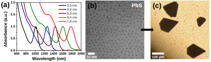
Fig. 1: (a) Absorption spectra of in-house synthesized PbS NCs with different sizes, (b) TEM image of PbS NCs with diameter of 3.5 nm,
(c) optical micrograph of three-dimensional supercrystals self-assembled from particles shown in (b).
The research of our group focuses on the investigation of structure and dynamics of various colloidal nanoparticles by means of X-ray scattering techniques, i.e. small angle X-ray scattering (SAXS), wide angle X-ray scattering (WAXS)), X-Ray photon correlation spectroscopy (XPCS) and X-ray cross correlation analysis (XCCA) .
The in-house synthesis includes a broad range of nanomaterials, such as semiconductor, metal, dielectric, magnetic, polymeric particles of different sizes and shapes. The synthesis is carried out by wet chemical methods and results in nanostructures with a narrow size and shape distribution. In order to explore their detailed properties, the nanostructures are characterized using microscopic, spectroscopic and/or scattering techniques.
Some examples of in-house synthesized colloidal particles are represented below.
Semiconductor Nanocrystals
|
Monodisperse semiconductor nanocrystals (e.g. lead sulfide) are synthesized in a colloidal solution using Schlenk line technique. The size of nanoparticles is controlled by the choice of precursors, ligands, solvents and reaction conditions (concentration, growth time, injection and growth temperature, etc.). Lead sulfide exhibits extensive quantum-size effects in nanocrystalline form and possesses tunable optical properties. Size-controlled synthesis of high quality PbS nanocrystals (NCs) is usually carried out by a hot-injection method. By this technique, the bandgap of PbS NCs can be tuned from 1.4 to 0.7 eV (absorption maximum from 900 to 1800 nm) by changing the size from 3 to 8 nm. Figure 1a shows normalized absorption spectra of in-house synthesized PbS NCs with different sizes. Due to their high degree of monodispersity (Fig. 1b), PbS NCs self-assemble into 3D supercrystals on a substrate during slow solution destabilization by a non-solvent (Fig. 1c). |
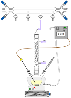
Fig. 2: Synthesis of semiconductor nanocrystals. |
Gold Nanoparticles
Besides semiconductor nanocrystals our group produces metal nanoparticles and nanorods, which are interesting for a wide range of potential technological applications.
Gold nanorods (NRs) have recieved significant scientific attention due to their tunable longnitudinal surface plasmon resonance (LSPR) across a broad spectral range. The scattering and absoprtion cross sections at a given LSPR wavelength are largely determined by the overall size of Au NRs, which is why the uniformity of size and shape becomes highly important.
Typical TEM image of in-house synthesized Au NRs without any post-synthetic size selection shows monodisperse sample (Fig. 3 (left)). The wavelength of the LSPR band is tuned by controlling the aspect ratio (length-to-diameter) of the gold nanorod (Fig. 3 (right)).
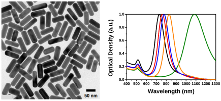
Fig. 3: TEM image of highly monodisperse gold nanorods (left) and UV-vis-NIR spectra of gold nanorods with different aspect ratio (right).
Silica Nanoparticles
TEM images of in-house synthesized colloidal silica particles and core-shell silica/poly-N-isopropylacrylamide (PNIPAM) spheres are shown below in Fig. 4. PNIPAM is a stimuli-responsive hydrogel that undergoes a sharp volume phase transition at a lower critical solution temperature in water around 32°C, which results in a rapid collapse of the particle radius. The size of the silica core and the PNIPAM shell is precisely controlled in a wet chemical synthesis. X-ray scattering techniques allow for the investigation of structure and dynamics of highly concentrated colloidal SiO2/PNIPAM solutions due to the strong scattering from the silica core.
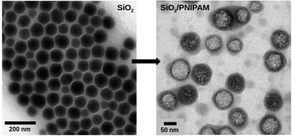
Fig. 4: TEM images of colloidal silica particles (left) and silica/PNIPAM core/shell particles (right).
Hematite Nanoparticles
Furthermore, monodisperse magnetic spindle-shaped hematite particles are synthesized in our laboratory with different aspect ratios. These nanospindles are involved in various X-ray scattering experiments, including investigation of their orientation in a liquid jet.

Fig. 5: TEM images of spindle-shaped hematite particles.